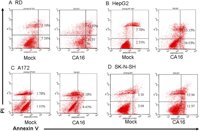
Figure 4. Membrane changes associated with apoptosis in CA16-infected cells.
(A) RD, (B) HepG2, (C) A172 and (D) SK-N-SH cells were inoculated with CA16 SHZH05 virus at the MOI of 1.0, 2.0, 1.0 and 1.0, respectively, or DMEM as a negative control for 24 h. Cells were washed with PBS, incubated with FITC-labeled Annexin V and stained with PI, followed by analysis via flow cytometry. Annexin V-positive/PI-negative cells were considered to be in early phase apoptosis. Annexin V-positive/PI-positive cells were considered to be in late phase apoptosis. Representative images are shown of three individual experiments (n = 3) performed for each cell line. *P<0.05.

