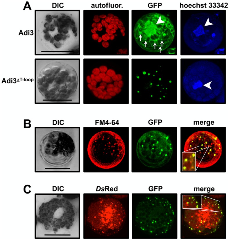
Figure 1. Deletion of the T-loop extension localizes Adi3 to the endomembrane system. Protoplasts of PtoR tomato plants were transformed with GFP-Adi3 constructs, protein expressed for 16 hrs, and the GFP signal viewed by confocal microscopy.
Compiled Z-axis images are shown. A, Localization of GFP-Adi3 proteins in tomato protoplasts. Protoplasts expressing the indicated GFP-Adi3 proteins were stained with Hoechst 33342 to label nuclear DNA (white arrow head). Small arrows indicate localization of punctate structures for GFP-Adi3 expression. B, GFP-Adi3ΔT-loop colocalizes with the endosomal vesicle stain FM4-64. Cells expressing GFP-Adi3ΔT-loop were treated with 20 µM FM4-64 for 2 hrs before visualization. Inlay shows zoom of region of interest. C, GFP-Adi3ΔT-loop colocalizes with the endosomal vesicle marker 2xFYVE-DsRed. GFP-Adi3 ΔT-loop and 2xFYVE-DsRed constructs were cotransformed into protoplasts and expressed for 16 hrs before visualization. Inlay shows zoom of region of interest. Bars = 20 µm.

