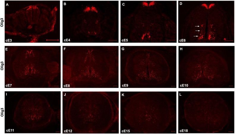
Figure 1. Developmental expression of Olig3 in the embryonic chick spinal cords at the thoracic level.
Cross sections from various embryonic chick spinal cord tissues were subjected to immunofluorescent staining with rat anti-Olig3 antibody. (A) cE3. (B) cE4. (C) cE5. (D) cE6. (E) cE7. (F) cE8. (F) cE9. (G) cE10. (H) cE11. (I) cE12. (J) cE15. (K) cE18. The dorsal part is up. Bars, 100 µm.

