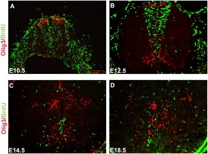
Figure 3. Short-term BrdU birth-dating analysis of Olig3+ cells in the developing mouse spinal cords.
Mouse embryos from E10.5 (A), E12.5 (B), E14.5 (C), and E18.5 (D) were pulse-labeled with BrdU for 2 hours. Transverse spinal cord sections were subjected to double immunolabeling with anti-BrdU (green) and anti-Olig3 (red). Many Olig3/BrdU double-positive cells were observed at the dorsal-most domain of spinal cord at E10.5. In contrast, no Olig3/BrdU double-positive cells could be found at E12.5, E14.5 and E18.5. The dorsal part is up. Bars, 100 µm.

