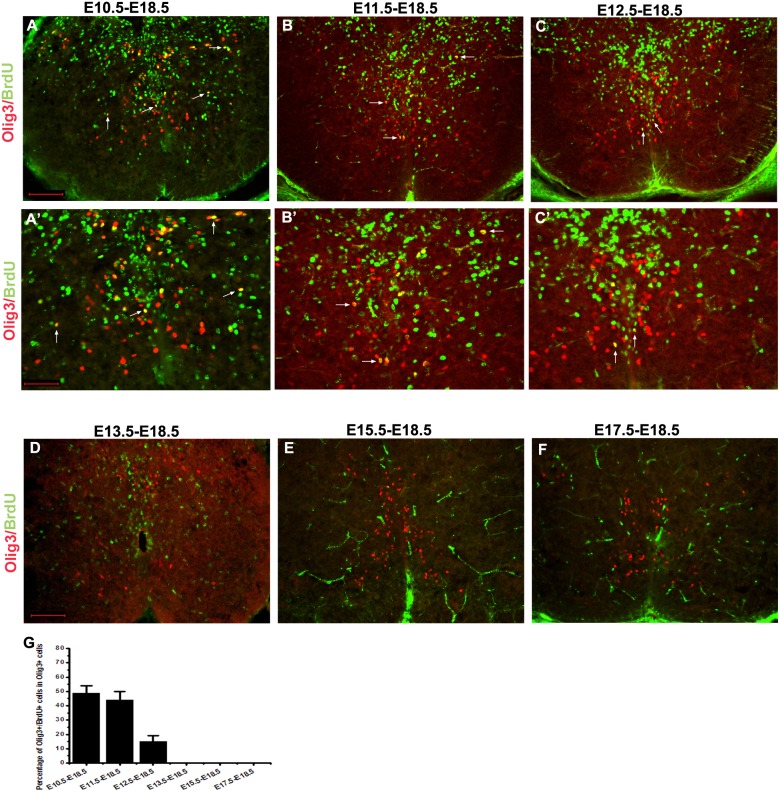
Figure 4. Long-term BrdU birth-dating analysis of Olig3+ cells in the developing mouse spinal cords.
BrdU was injected into pregnant mice at various embryonic stages and embryos were harvested at E18.5. Transverse spinal cord sections were subjected to double immunolabeling with anti-BrdU (green) and anti-Olig3 (red). Spinal cord section from mice injected at E10.5, E11.5 and E12.5 contained BrdU+/Olig3+ cells. However, injection after E13.5 did not produce any Olig3/BrdU double-positive cells. A’–C’ are the high-magnification images of A–C, respectively. Double positive cells are indicated with arrows. (G) The percentage of Olig3+ cells that co-express BrdU was calculated. The dorsal part is up. Bars: A–F are 100 µm; A’–C’ are 50 µm.

