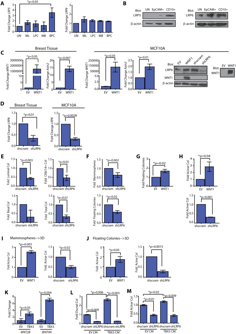
Figure 2. WNT enhances acinar progenitor activity through LRP6 signaling.
(A) LRP5 expression was enriched in basal progenitor cells (BPC; EpCAM−CD10+), while LRP6 expression was present at similar levels in all fractions. Mammary epithelial cells (MECs) were sorted and expression levels were quantified by qPCR (n = 6 patient samples; mean±s.d.). UN = unsorted, ML = EpCAM+CD49f−, LPC = EpCAM+CD49f+, MB = EpCAM+CD10+. (B) LRP6 protein was detected in cell lysates from EpCAM+ luminal cells and CD10+ basal cells, while LRP5 protein was detected in lysates from CD10+ basal cells. (C) WNT1 overexpressing MECs (n = 3 patient samples) and MCF10A cells (n = 3 experiments) demonstrated significantly elevated expression of WNT1 and downstream target, axin2. Expression was analyzed using qPCR (mean±s.d.). WNT1 protein was detected in cell lysates and conditioned media from WNT1 MCF10A cells. Protein for LRP6 was decreased in shLRP6 MCF10A cells compared with shscrambled (shscram) cells. Representative images from 3 experiments. (D) LRP6 expression was significantly reduced in MECs (n = 3 patient samples) and MCF10A cells (n = 3 experiments) transduced with shLRP6 lentivirus compared with those transduced with shscram. Expression was analyzed using qPCR (mean±s.d). (E) shLRP6 MECs demonstrated significantly reduced colony formation on adherent plates compared with shscram cells (n = 3 patient samples; mean±s.e.m). (F) shLRP6 MECs demonstrated reduced mammospheres and floating colony formation compared with controls (n = 4 patient samples; mean±s.e.m). (G) WNT1 MECs demonstrated significantly increased floating colony formation (n = 6 patient samples; mean±s.e.m.). (H) WNT1 MECs significantly increased acinar colony formation, while shLRP6 MECs significantly decreased acinar colony formation (n = 6 patient samples; mean±s.e.m.). (I, J) WNT1 expression increased acinar colonies, while reduced LRP6 expression diminished acinar colonies, when WNT1, EV, shscram, and shLRP6 expressing MECs were grown as mammospheres (I) or floating colonies (J) prior to plating on collagen (n = 5 patient samples; mean±s.e.m.). (K) TBX3 MCF10A cells demonstrated significantly increased expression of WNT2B and WNT9A compared with EV cells. Expression levels were quantified using qPCR and denoted as fold change compared to control cells (n = 3 experiments; mean±s.d.). (L) Ductal colonies were significantly increased in shscram MCF10A cells exposed to conditioned media (CM) isolated from TBX3 MCF10A cells compared to those grown in CM from EV MCF10A cells. Exposure to TBX3 CM did not enhance ductal colonies in shLRP6 MCF10A cells (n = 3 experiments; mean±s.e.m.). (M) shscram MCF10A cells demonstrated significantly increased acinar colony formation when treated with CM from TBX3 MCF10A cells compared to CM from EV MCF10A cells. Exposure to TBX3 CM did not enhance acinar colonies in shLRP6 MCF10A cells (n = 3 experiments; mean±s.e.m.).

