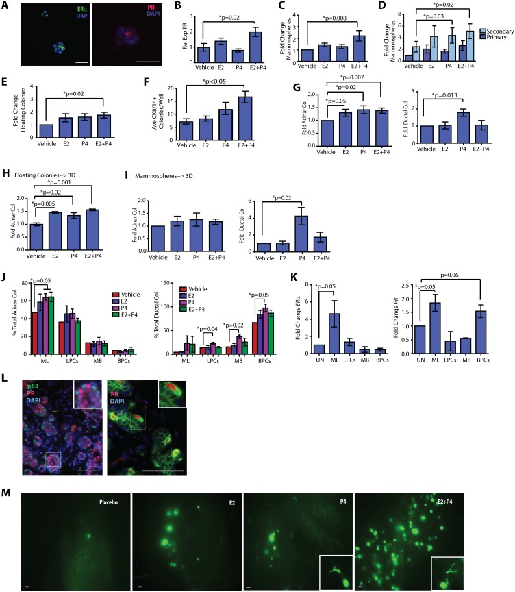
Figure 3. Progesterone increases basal ductal progenitor activity.
(A) Mammospheres formed from mammary epithelial cells (MECs) expressed estrogen receptor alpha (ERα) and progesterone (PR) receptor. (B) Mammospheres treated with E2+P4 significantly upregulated PR expression. PR expression was detected by qPCR (n = 3 patient samples; mean±s.d.). (C) 17β-estradiol (E2) + progesterone (P4) treatment significantly enhanced MEC mammosphere numbers (n = 9 patient samples; mean±s.e.m.). (D) P4 and E2+P4 treatment significantly enhanced secondary mammosphere numbers (n = 6 patient samples; mean±s.e.m.). (E) E2+P4 treatment of MECs significantly increased floating colony growth (n = 9 patient samples; mean±s.e.m.). (F) E2+P4 treatment of MECs significantly enhanced the growth of bi-potent (CK8/14+) colonies in adherent culture (n = 3 patient samples; mean±s.e.m.). (G) On collagen, E2, P4, and E2+P4 significantly increased acinar colonies. P4 significantly enhanced ductal colonies (n = 9 patient samples; mean±s.e.m.). (H) E2, P4, and E2+P4 significantly increased acinar colonies. MECs were grown as floating colonies, then plated with hormones on collagen (n = 3 patient samples; mean±s.e.m.). (I) P4 significantly increased ductal colonies. MECs were grown as mammospheres, then plated on collagen with hormones (n = 7 patient samples; mean±s.e.m.). (J) P4 and E+P significantly increased acinar colonies in mature luminal (ML; EpCAM+CD49f−) cells, and P4 significantly increased ductal colonies in luminal progenitor cells (LPC; EpCAM+CD49f+), mature basal (MB; EpCAM+CD10+), and basal progenitor cells (BPC; EpCAM−CD10+). MECs were sorted and grown on collagen in the presence of hormones (n = 3 patient samples; mean±s.e.m.). Data are represented as a fold change compared to the vehicle control for each cellular population multiplied by the percentage of total acinar or ductal colonies formed. (K) ERα expression was enriched in ML cells; PR expression was enriched in ML and BPCs. MECs were sorted and qPCR performed (n = 6 patient samples; mean±s.d.). (L) PR expression was present in the basal epithelial layer adjacent to p63 expressing epithelial cells (inset). Basally located PR expressing cells also expressed CK8 (inset). (M) Primary epithelial cells (MEC) were isolated from reduction mammoplasty tissues, transduced with GFP lentivirus, and grown in the humanized fat pads of ovariectomized NOD/SCID mice treated with E2, P4, E2+P4, or placebo pellets. Glands from P4 and E2+P4 treated mice demonstrated increased growth of ductal structures (inset; n = 3 experiments). Scale bars = 100 µm.

