
Fig. 5.
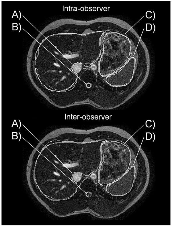
Top: Example of intraobserver contouring trials. (A) Differences in extension of liver contour around the vena cava lead to large (poor) HD values. (B and D) Great contouring precision of the spinal cord (black) and spleen (white). (C) Good contouring precision of the stomach. Bottom: Example of interobserver contouring trials. (A) Differences in extension of liver contour around the vena cava lead to very large (poor) HD values. (B) Small slicewise differences in contours of the spinal cord (black) lead to low (poor) DC values when summed over all slices. (C) Good contouring precision of the stomach. (D) Great contouring precision of the spleen.