
Figure 3.
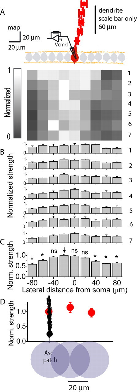
High-resolution mapping confirms the relative homogeneity of the granule cell population. A, The average normalized EPSC map and dendritic orientation (red) obtained from six Purkinje cells from experiments similar to that presented in Figure 2. B, A row-by-row analysis of the average normalized EPSC map shown in A. Each row was normalized to the largest value in that row. C, A column-by-column analysis of the average normalized EPSC map shown in A. Shown is the normalized average of the seven rows shown in A. * denotes statistical significance with respect to the column denoted by the arrow at p < 0.005. D, For each of the EPSC mapping experiments averaged in A above, two investigators independently determined the location of the “ascending” patch in the row closest to the Purkinje cell layer. The average peak EPSC amplitude of the position that corresponded to the location where granule cells form ascending synapses (Asc patch) was taken as one. The relative peak EPSC amplitudes of the adjoining patches located on either side of the ascending patch and the second adjacent pair located 40 μm away from the ascending patch were pooled and averaged. Data are presented as mean ± SEM.