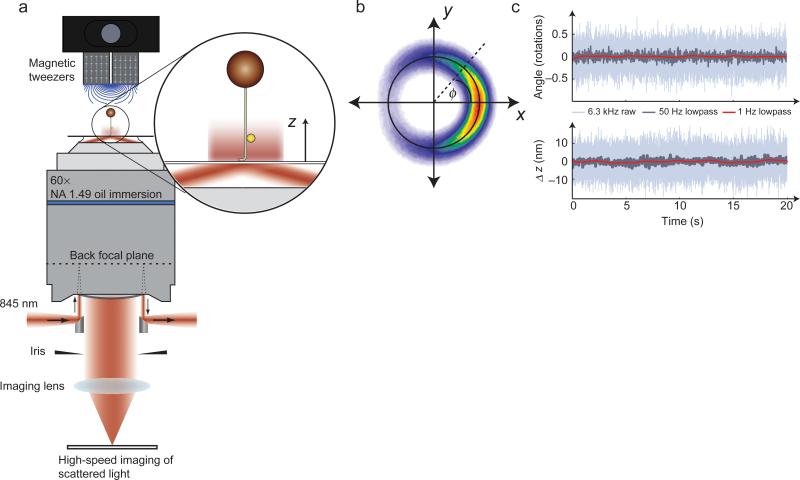
Figure 1. AuRBT design and data collection.
(a) Combining 3D nanoparticle tracking with magnetic tweezers. An excitation laser is introduced using a small 45-degree mirror to achieve total internal reflection. The return beam is extracted using a second mirror, and stray light is rejected with an iris. The darkfield image of a gold rotor bead is imaged on a high-speed camera. Evanescent illumination excludes the larger magnetic bead from the excitation field, while providing an intensity gradient for tracking z displacements. (b) 2D tracking (see also Supplementary Video 1). A rotor bead was attached above a 420 bp torsionally constrained DNA segment and held under 10 pN of tension. The XY position was recorded at 6.3 kHz and accumulated over 150 s to generate a 2D histogram, which shows restriction to an annulus and a preferred equilibrium angle. The instantaneous in-plane angle φ can be calculated from the x and y measurements. The measured diameter of the orbit for this bead (black circle) is d = 69.4 nm, which is a measure of the size of the particle. (c) Simultaneous angle and extension measurements of a rotor (d = 68.1 nm) attached above a 420 bp torsionally constrained segment and held under 26 pN of tension by a magnetic bead dimer. Extension was measured using evanescent nanometry (see Methods and Supplementary Fig. 3). Full-bandwidth traces are shown along with 50 Hz and 1 Hz lowpass filtered data.

