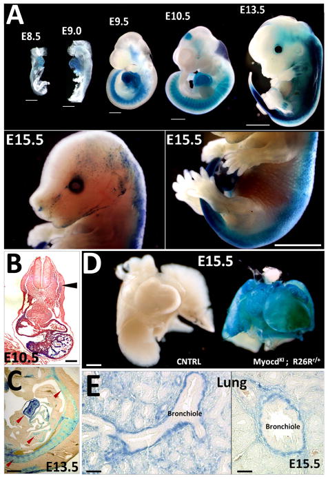
Figure 3. Myocardin expression pattern in MyocdKI/+; R26Rr/+ reporter mice.
A) X-gal staining at different stages of developing mouse embryos. LacZ expression is observed in the developing heart, somites and mid-brain. Note that LacZ expression expands to proximal limb internal structures (from E13.5) (Scale bars, 250μm). B) Transversal section of an E10.5 embryo showing cardiac specific expression of LacZ, somite expression is low compared to the heart (arrowhead) (Scale bar, 500μm). C) X-gal staining on a sagital section of an E13.5 embryo reveals LacZ expression in the heart, somite derivatives and forming vasculature (arrowhead) (Scale bar, 500μm). D) LacZ staining in an E15.5 MyocdKI/+;R26Rr/+ embryo heart and lung. A wild type littermate embryo was used as a control (Scale bar, 500μm). E) Cross sections of a MyocdKI/+;R26Rr/+ E15.5 embryo lung after X-gal staining showing LacZ expression in the smooth muscle layer surrounding the forming bronchioles (Scale bars, 125μm).

