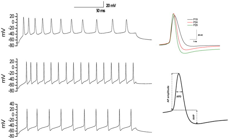
Fig. 1. Typical recordings of evoked APs by depolarizing current injection in DCN neurons.

Left panel (Top, middle, and bottom) represents typical whole-cell current clamp recordings with depolarizing current injection of 0.1nA in DCN neurons from P19, P24, and P29 rats, respectively. Right top panel represents the corresponding APs. Right bottom panel represents AP measures taken.
