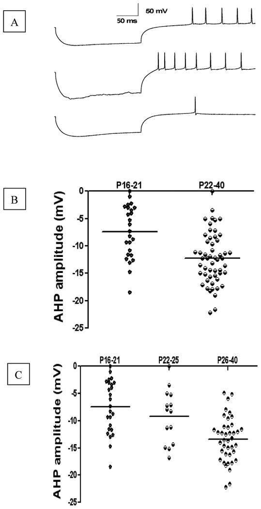
Fig. 4. Developmental changes in AHP amplitude of DCN rebound spikes elicited by hyperpolarization pulses.

A represents typical rebound spikes elicited by hyperpolarization current injection of -0.5nA in DCN neurons from P19, P24, and P29 rats, respectively. B represents AHP amplitude of rebound spikes from P16-21 group, and P22-40 group, respectively. C represents AHP amplitude of rebound spikes from P16-21 group, P22-25 group, and P26-40 group, respectively. Note that each point in the graphs represents the result from a single neuron, and dash line illustrates the average at a given age. The developmental changes happened on P22-25 (weaning age).
