Figure 2.
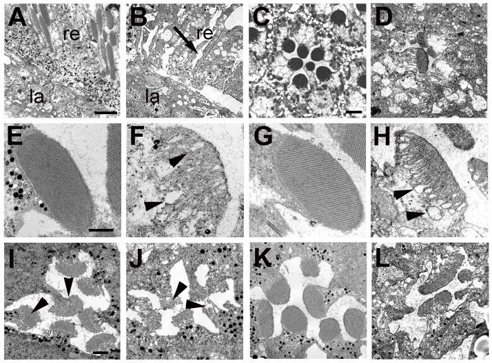
Transmission electron microscopy reveals degeneration of rhabdomeres. A: A GMR-GAL4/UAS-LacZ fly at 36 hours post eclosion shows an intact retina (re) and lamina (la). B: In a 36hr old GMR-GAL4; tauGD25023 fly the retina is disorganized and vacuoles have formed in the retina (re, arrow). C: An intact ommatidia in a 36h old GMR-GAL4/UAS-LacZ control with seven photoreceptors present. D: In contrast, a 36h old GMR-GAL4; tauGD25023 fly shows a disrupted ommatidial structure with only a few abnormal looking photoreceptors present. Whereas control flies have highly structured rhabdomeres at this age (E), the rhabdomeres of the photoreceptors that are present in GMR-GAL4; tauGD25023 have an abnormally loose structure with loops forming that are mostly found in proximity to the cell cytoplasm (F, arrowheads). G: In contrast, the rhabdomeres are intact in a 1d old tauDf(3R)MR22/Df(3R)BSC499 fly but become more loose when aged for 7d (H). I: P11 control pupa showing the developing seven rhabdomeres (arrowheads). J: In an age-matched GMR-GAL4; tauGD25023 fly, the rhabdomeres already look abnormal although all photoreceptors are present at this stage. K: Pharate adult control fly. L: A pharate adult GMR-GAL4; tauGD25023 fly has lost several photoreceptors and the remaining photoreceptors are abnormally shaped and have irregular rhabdomeres. Scale bars in A=6μm, in C=2μm, in E=1μm, and in I=1μm.

