
Figure 4.
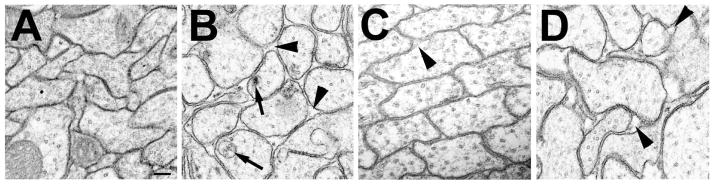
Decreasing the levels of dTau affects axonal morphology. A: Transmission electron microscopy reveals tightly packed axons in a 1d old wild type fly. B: In a 1d old the tauGD25023; elav-GAL4 fly the axons look more rounded and extracellular gaps are visible (arrowheads). In addition, intracellular inclusions and vacuoles can be detected (arrows). C: In tauDf(3R)MR22/Df(3R)BSC499, the axons are tighter packed and only a few extracellular gaps are visible (arrowhead) at 1d of age, however, the number and size of these gaps increases with age as seen in a 16d old fly (D). Scale bar in A=200μm.