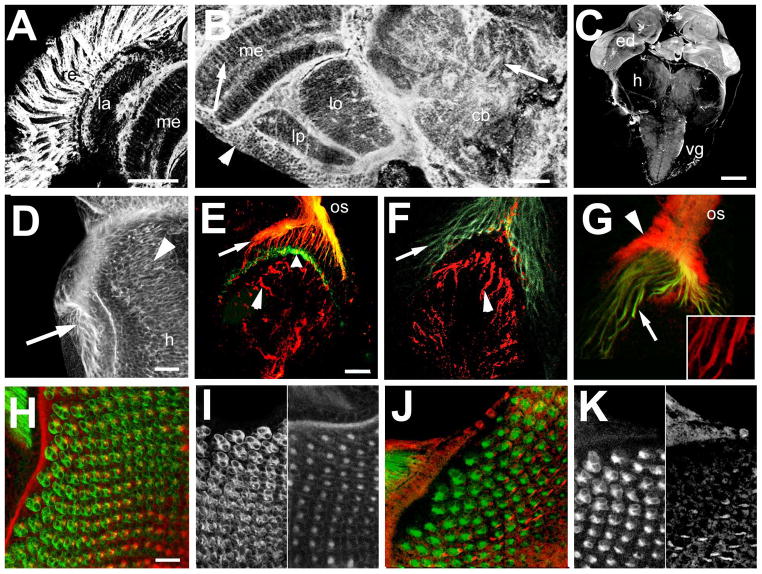
Figure 7.
dTau is expressed in the eye and brain. A, B: Vibratome sections stained with anti-dTau show that dTau is strongly expressed in the adult eye (A, confocal stack of 25 0.5μm optical sections) but it can also be found in the brain (B, confocal stack of 20 images). dTau is detectable in cell bodies localized in the cortex (arrowhead) and in neuronal fibers in the neuropils (arrows). C; In the larval brain, strongest expression is found in the eye discs (ed) but lower levels of expression are detectable in the proximal half of the hemispheres (h) and in the ventral ganglia (vg). D: Weak expression can also be detected in the outer optic anlagen (arrowhead, the arrow points to the photoreceptor axons). E: In wild type, dTau (red) co-localizes with chaoptin (24B10, green) in photoreceptor axons projections (arrow) but does not appear to be present in their terminals (broad arrowhead). It can also be seen in fibers in the developing lamina (arrowhead). F: In GMR-GAL4; tauGD25023, dTau is still present in the fibers in the lamina (arrowhead) but is not detectable in photoreceptor axons (arrow). G: In contrast, in tauDf(3R)MR22/Df(3R)BSC499 dTau is still present in photoreceptor axons, though at much lower levels than in wild type (arrows and inset). In addition, there is a strong staining in the optic stalk (os) that does not co-localize with 24B10 (arrowhead). H: dTau is also detectable in the eye disc (stack of 15 optical images of 0.5μm), co-localizing with 24B10. However, the dTau appears restricted to axons (I, right panel), whereas 24B10 also outlines the photoreceptor cell bodies (I, left panel). J, K: In eye discs from tauDf(3R)MR22/Df(3R)BSC499 flies, the pattern again looks different with a less distinct and punctuate dTau staining (K, right panel). Scale bar in A=50μm, in B=25μm, in C=100μm, in D=20μm, in E=25μm, and in H=20μm

