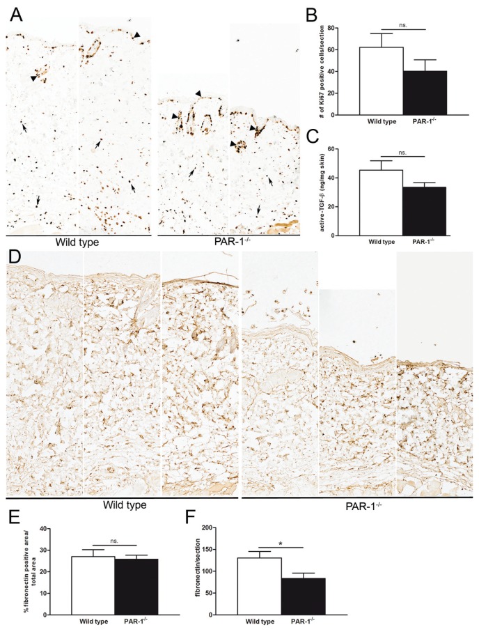
Figure 3.
Modification of profibrotic processes by PAR-1 in vivo. (A) Immunohistochemical staining of Ki67-positive cells in skin sections of wild-type and PAR-1-deficient mice upon bleomycin treatment (10× magnification). (B) Quantification of Ki67 positive cells in the dermis (excluding cells of hair follicles and sebaceous glands [arrow heads in panel A]) of wild-type and PAR-1-deficient mice upon bleomycin treatment (n = 4–7). (C) Active TGF-β levels in skin homogenates of wild-type and PAR-1-deficient mice upon bleomycin treatment (n = 7–8). (D) Representative pictures of fibronectin expression in skin sections from wild-type and PAR-1-deficient mice upon bleomycin treatment (10× magnification). (E) Density of fibronectin in the dermis of bleomycin-treated wild-type and PAR-1-deficient mice (n = 5–6). (F) Total amount of fibronectin per dermal section of bleomycin-treated wild-type and PAR-1-deficient mice. Data are means ± SEM. *p < 0.05. n.s., Not significant.

