
Figure 1.
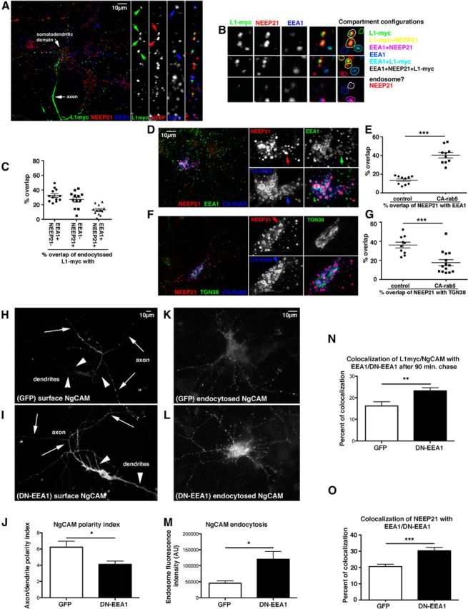
EEA1 interference leads to missorting of L1/NgCAM. A–C, EEA1-negative/NEEP21-positive compartments are endosomes. Neurons were transfected with L1-myc, and endocytosis assays were performed with anti-myc antibodies for 20 min. Endocytosed L1-myc was detected with secondary antibodies (green) and counterstained against NEEP21 (red) and EEA1 (blue). A, Overview of one neuron and zoom of a single dendrite. Single channels and triple merges are shown in the side panels. Examples of compartments are shown in detail in B. The overlap of markers is color coded on the right. C, The percentage of overlap of L1-myc with E+N+, E−/N+, and E+/N− endosomes was quantified. A scatter plot of individual cell counts is plotted, with means and SEM indicated. Twelve cells were analyzed. D–G, NEEP21 accumulates in enlarged endosomes induced by expression of constitutively active (CA)-rab5. Neurons were transfected with GFP-CA-rab5 (blue) or soluble GFP (control; data not shown) for 24 h, and then fixed and stained against NEEP21 (red) and EEA1 (D, green) or TGN38 (F, green). Single channels and a triple-stained merged image of one soma are shown enlarged in the side panels. Colocalization of NEEP21 with EEA1 (E) or TGN38 (G) was quantified and plotted in a scatter plot showing individual cells, as well as mean and SEM. Student's t test, p < 0.0001. Ten cells per condition were analyzed. H–J, EEA1 interference leads to missorting of surface NgCAM. Neurons were transfected with NgCAM and either GFP as control (H) or GFP-EEA1-DN (I) for 24 h, and then surface NgCAM was stained in nonpermeabilized cells after fixation. Arrows indicate axons, and arrowheads indicate dendrites. The average NgCAM surface staining intensity was determined for GFP-transfected cells (n = 24 cells) and GFP-DN-EEA1 (n = 38 cells) along the axon and dendrites, and the axon/dendrite polarity index was calculated (J). Bar indicates the SEM. *p < 0.01, Student's t test. One representative experiment is shown (from a total of three independent experiments). K–N, Endocytosed NgCAM accumulates in somatodendritic endosomes when EEA-DN is coexpressed. Neurons were transfected with NgCAM and either GFP as control (K) or GFP-EEA1-DN (L) for 24 h and anti-NgCAM antibody was fed to live cells for 20 min. Endocytosed NgCAM was detected with a secondary antibody after acid stripping of surface-bound antibody. The extent of endocytosed NgCAM in somatic endosomes was quantified (M). N = 47 cells (GFP), 51 cells (EEA1-DN). Bar indicates the SEM. *p < 0.01, Student's t test. One representative experiment is shown (from a total of three independent experiments). N, Endocytosed L1-myc accumulated significantly in EEA1-positive endosomes after 90 min of endocytosed L1-myc antibody chase. N = 21 cells (GFP), and N = 34 cells (DN-EEA1). Bar indicates the SEM. **p < 0.001 Student's t test. One representative experiment is shown (from a total of three independent experiments). O, Overexpression of GFP-EEA1-DN in neurons caused increased accumulation of NEEP21 in EEA1 endosomes. N = 17 cells (GFP), and N = 12 cells (DN-EEA1). Bar indicates the SEM. ***p < 0.0001, Student's t test.