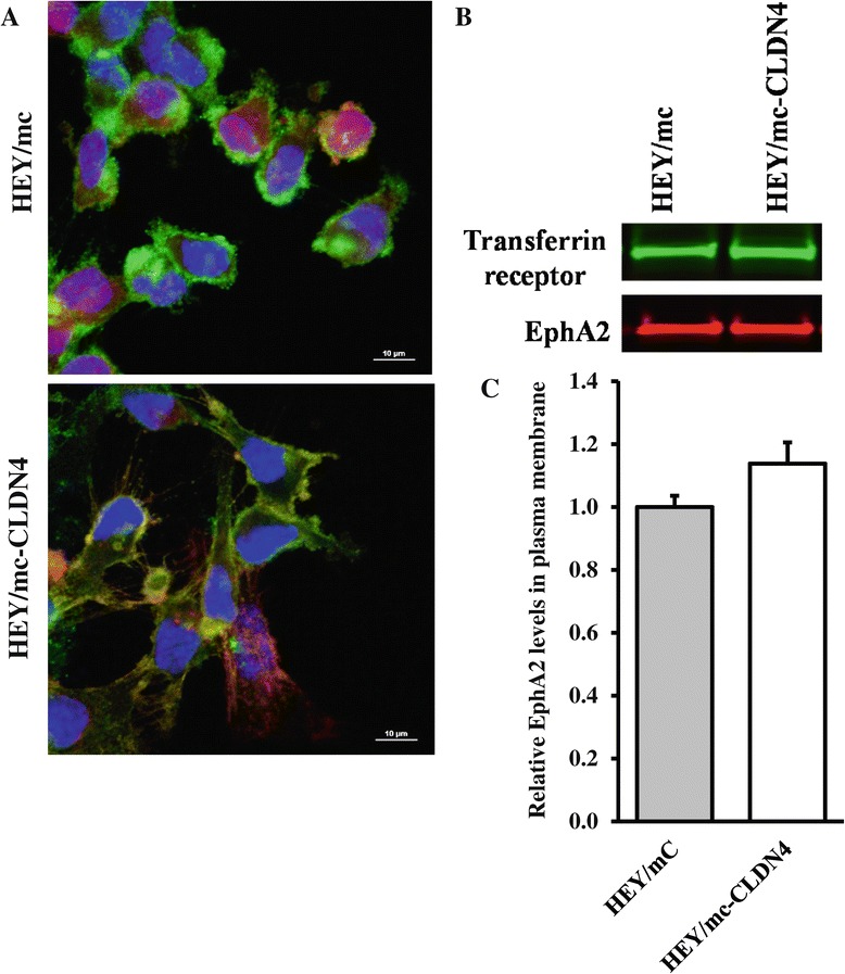
Figure 7.

Effect of CLDN4 on the subcellular localization of EphA2 in the absence of E-caderin. (A) Upper panel, HEY/mc cells expressing mCherry (red) stained with anti-EphA2 antibody (green). Lower panel, HEY/mc-CLDN4 cells expressing mCherry fused to CLDN4 stained with anti-EphA2 antibody (green). Cell nuclei were counterstained with DAPI (blue). Yellow staining indicates co-localization. (B) EphA2 levels in the plasma membrane of HEY/mC and HEY/mc-CLDN4 cells as determined by Western blot analysis of biotinylated surface proteins. (C) Histogram summarizing the results of 3 independent Western blots. Data presented are mean ± SEM (n = 3).
