
Figure 10.
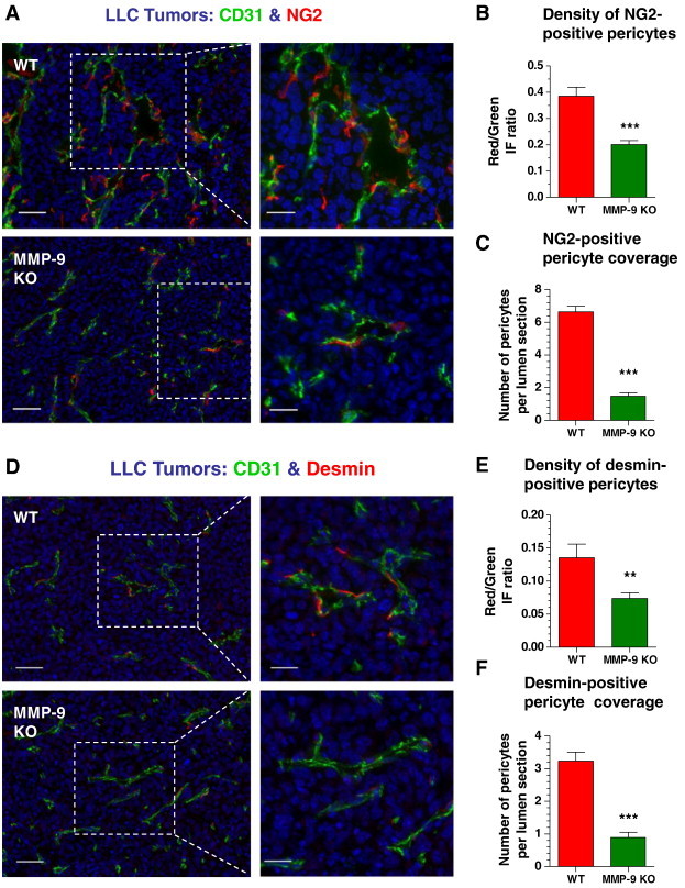
Microarchitecture of angiogenic blood vessels is supported by pericytes in an MMP-9–dependent manner. (A and D) WT and MMP-9 KO LLC tumors were stained for CD31 (green) and either NG2 (A) or desmin (D) pericyte marker (red). Bars, 50 μm (left) and 25 μm (right). (B and E) Pericyte density in LLC tumors grown in WT or Mmp9-KO mice was quantified in individual tumor sections as the ratio of IF signal from pericytes (red) versus IF signal from CD31-positive vasculature (green). A total of 34 to 38 tumor sections were analyzed in five WT and six Mmp9-KO tumors harvested in two independent experiments. ***P< .0001. (C and F) Pericyte coverage was determined as the number of NG2 (C) or desmin (F) positive cells per individual cross-sectioned lumen. A total of 31 and 29 (C) and 56 and 64 (F) vessels were analyzed in WT and Mmp9-KO tumors, respectively. ***P < .0001.