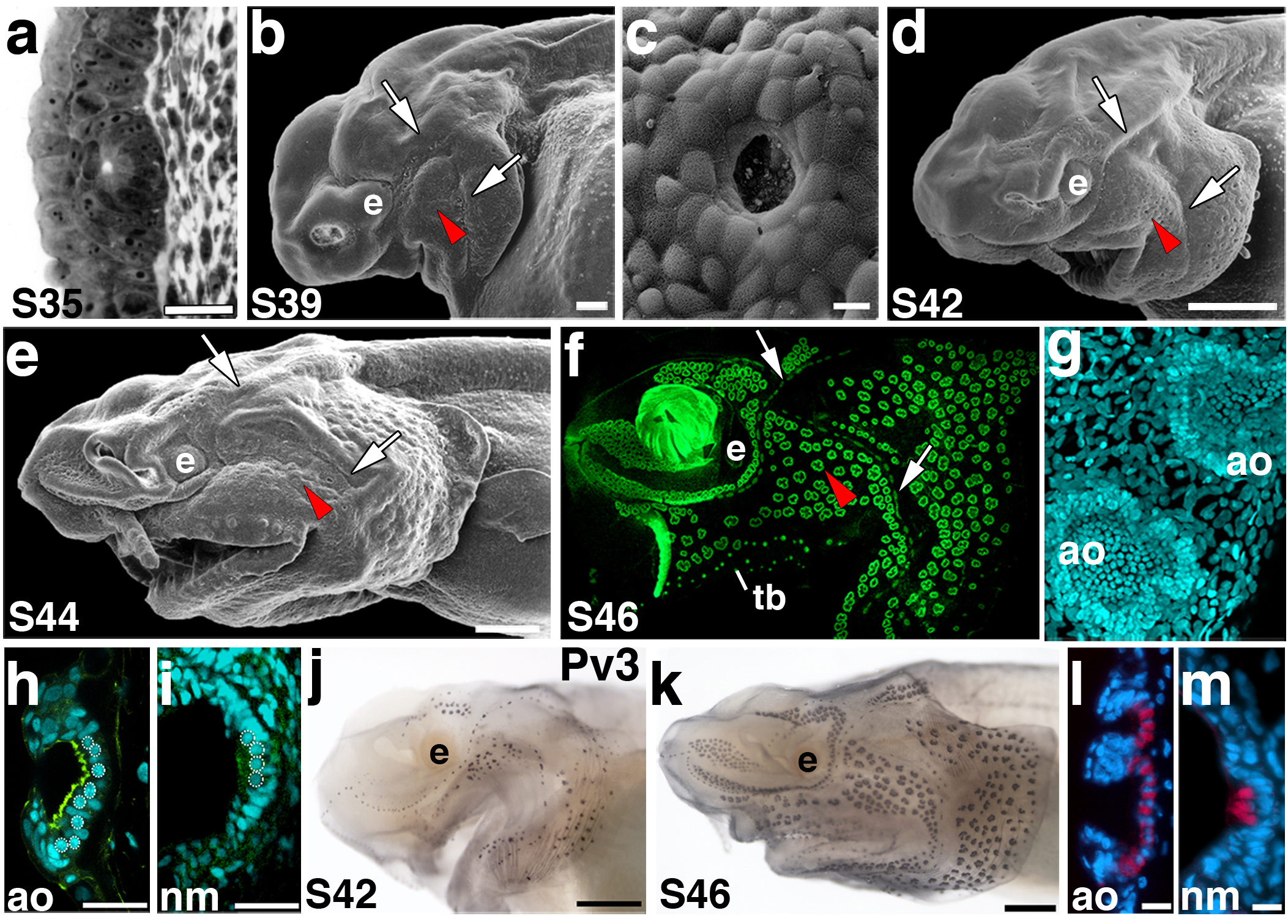
Figure 3. Ampullary organ development.

Anterior to left; lateral views unless otherwise noted. (a) Transverse section through an ampullary organ primordium at stage 35. (b) SEM of a stage 39 embryo. Ampullary organs have erupted through the surface in several fields, in particular on the opercular flap. The red arrowheads in panels b-f indicate approximately the same region of ampullary organs that are erupting or have erupted at the different stages of development. The arrows indicate neuromast canal lines, specifically of the otic and preopercular lines. (c) SEM showing a surface view through the pore of a newly erupted ampullary organ. (d) SEM of a stage 42 embryo. Ampullary organs appear to have erupted from all fields by this stage. (e) SEM of a stage 44 embryo showing further differentiation of the ampullary organ fields. (f) Fluorescent image of a stage 46 embryo skin mount, stained with the nuclear marker Sytox Green. Individual clusters of ampullary organs are discernable, as are the neuromast canal lines. (g,h) Confocal images of ampullary organs stained with the nuclear marker DAPI in (g) surface view and (h) transverse view. The F-actin marker phalloidin (green) labels the microvilli-enriched apical surface. (i) Confocal image of a neuromast in transverse view at the same stage for comparison with h. In h and i, the round nuclei of sensory cells are outlined. (j) Stage 42 embryo stained for parvalbumin-3 (Pv3). Hair cells within both neuromasts and ampullary organs express this Ca2+-binding protein. (k) Stage 46 embryo stained for parvalbumin-3. Pv3 is strongly expressed in all lateral line sensory organs. (l, m) False colour overlay with DAPI images, respectively, of transverse sections through ampullary organs and a neuromast. Pv-3 (red in l, m) specifically labels the sensory hair cells in both ampullary organs and neuromasts. Scale bars: 25 μm (a), 100 μm (b), 10 μm (c, l, m), 500 μm (d, e, j, k), 30 μm (h, i). Abbreviations: ao, ampullary organ; e, eye; S, stage; tb, taste bud.
