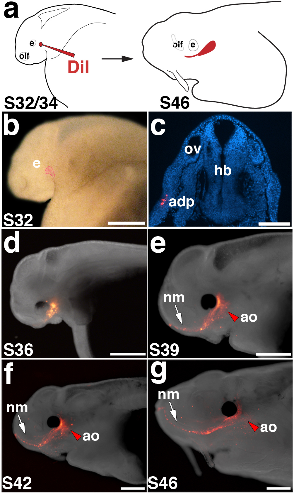
Figure 5. In vivo labelling of lateral line placodes.

(a) Schematic of in vivo labelling experimental design. Embryos are focally injected with DiI between stage 32 and 34 and followed to stage 46. (b) Stage 32 embryo immediately after focal injection with DiI. In this example, the anterodorsal placode (outlined in red) was labelled. (c) Transverse section through another stage 32 embryo immediately after injection, showing DiI-positive cells (red; DAPI in blue) in the anterodorsal placode. (d-g) Fluorescent images superimposed on darkfield images of the same embryo shown in panel b, at successive stages of development. (d) Stage 36. (e) Stage 39. (f) Stage 42. (g) Stage 46. As development progresses, DiI spreads anteriorly from the site of injection as neuromasts of the infraorbital line (arrow) are deposited. Ampullary organs (red arrowhead) are also labelled near the site of injection. Scale bars: 500 μm (b), 100 μm (c), 1 mm (d-g). Abbreviations: adp, anterodorsal lateral line placode; e, eye; hb, hindbrain; olf, olfactory; ov, otic vesicle; S, stage.
