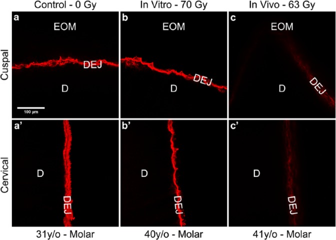
Figure 4.
Immunoreactivity for type IV collagen along the DEJ was reduced after in vivo high-dose radiotherapy. Immunofluorescently stained images were obtained by confocal microscopy as described in ‘Methods’. Results depicted are representative of 13 control (a, a′), 3 in vitro (b, b′), and 5 in vivo (c, c′) irradiated teeth, respectively. The immunostained appearance of the cuspal (a-b) and cervical (a′-b′) regions of control tooth sections (0 Gy) and in vitro-irradiated tooth sections (70 Gy) was consistently more robust than that for cuspal (c) and cervical (c′) regions from age-related in vivo-irradiated (> 60 Gy) teeth. EOM, enamel organic matrix; DEJ, dentin-enamel junction; D, dentin. Scale bar = 100 μm, applicable to all images.

