Figure 1.
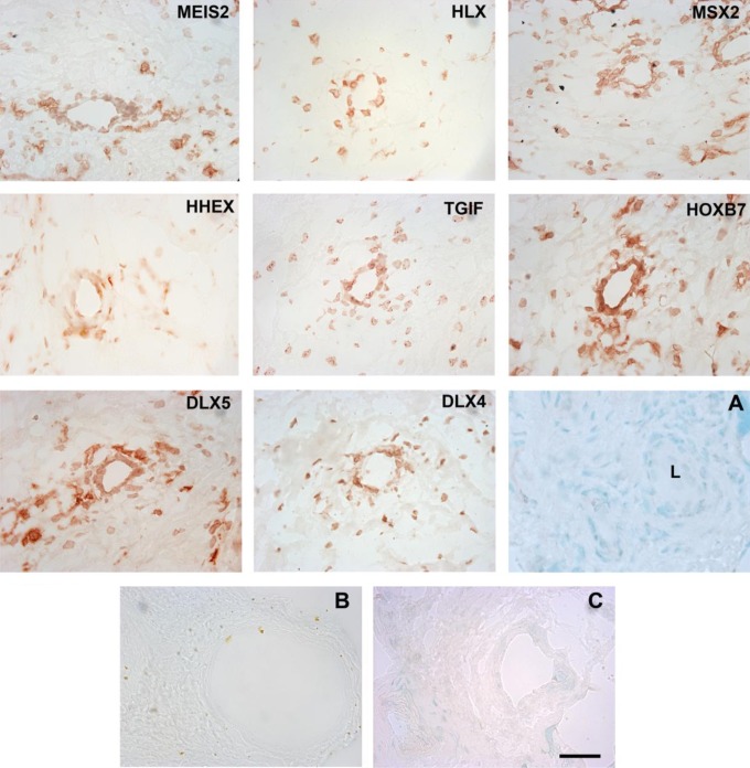
Immunohistochemical staining of first trimester human placental tissue (n = 3) with antibodies to MEIS2, HLX, MSX2, HHEX, TGIF, HOXB7, DLX5, and DLX4. Panels are labeled with the antibodies to homeobox gene protein products, which were used to generate the staining pattern. Panel A is a methyl green nuclear stain showing a ∼50 μm vessel and surrounding stromal morphology. L indicates lumen. Panels B and C show representative controls. Panel B shows an IgG1 control for mouse monoclonal antibodies (MEIS2, MSX2, and TGIF), the IgG2 control for mouse monoclonal antibodies (DLX5) showed a similar low background level of staining (data not shown). Panel C shows a preimmune serum control for rabbit polyclonal antibodies (HLX, HHEX, and DLX4). Detection was with AEC. Magnification was ×400 and the scale bar is 50 μm. IgG indicates immunoglobulin. MEIS2 indicates Meis1, myeloid ectropic viral integration site 1 homolog 2; HLX, H2.0-like Drosophila; MSX2, Msh homeobox 2; HHEX, hematopoietically expressed homeobox; TGIF, transforming growth factor β-induced factor; HOXB7, homeobox B7; DLX5, distal-less homeobox 5; DLX4, distal-less homeobox 4. (The color version of this figure is available in the online version at http://rs.sagepub.com/.)

