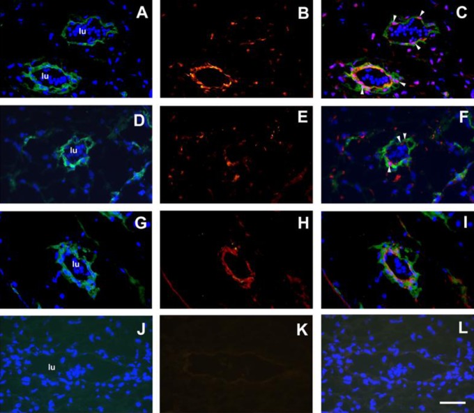
Figure 2.
Multilabel immunofluorescence staining of first trimester human placental stroma and blood vessels with DLX5 and HLX antibodies (n = 3). Panel A, FZD9 staining (donkey antimouse Alexafluor488, green) and DAPI staining (blue). Panel B, DLX5 staining (goat antirabbit Alexafluor568, red). Panel C, Combined FZD9/DAPI/DLX5 image. Panel D, FZD9 staining and DAPI staining. Panel E, HLX staining (red). Panel F, combined FZD9/DAPI/HLX image. White arrowheads show representative cells that stain positively with DLX5, DAPI (purple nuclei), and FZD9 (Panel C), or with HLX, DAPI (purple nuclei), and FZD9 (Panel F). Panel G, FZD9 staining and DAPI staining. Panel H, ITGA1 staining (red). Panel I, Combined FZD9/DAPI/ITGA1 image. Panel J, Mouse monoclonal X63 (green) and DAPI. Panel K, nonimmune rabbit serum (NIRS; red). Panel I, Combined X63/DAPI/NIRS image. Nucleated erythrocytes were detected within the vessel lumen (lu) by DAPI staining (Panels A, D, G, and J). Magnification was at ×600 and scale bar is 50 μm. DAPI indicates 4′,6-diamidino-2-phenylindole. HLX indicates H2.0-like Drosophila; DLX5, distal-less homeobox 5.

