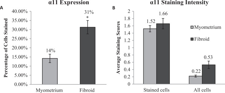
Figure 3.
Quantification of α11 staining in myometrium and fibroid samples. Of 10 195 myometrial cells, 1427 (14%) stained positive for α11, while 1453 of 4 686 fibroid cells (31%) stained positive (A); *p-value < 0.05. The intensity of the stained cells was similar in both myometrium and fibroid, and while the average intensity of all cells in each sample trended towards greater intensity in fibroid samples, this was not statistically significant (B).

