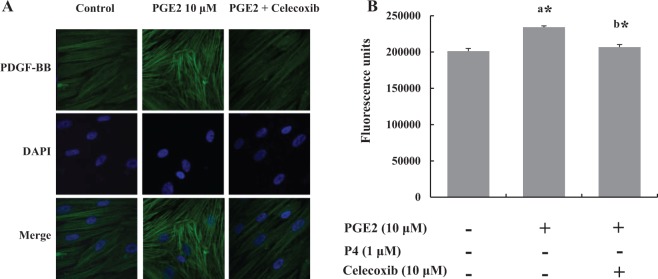
Figure 5.

Leiomyoma cells (n = 5) were treated with prostaglandin E2 (PGE2) or PGE2 plus celecoxib for 3 days. A, Representative fluorescence microscopy visualization showed the effect of celecoxib on PGE2-stimulated platelet-derived growth factor (PDGF) expression. The top panel shows representative images of the expression of PDGF, the middle panel shows corresponding DAPI nuclear staining, and the bottom panel shows merged images. B, Quantification of PGE2-stimulated fluorescence. The bars represent the mean ± standard error of values from triplicate experiments. a* indicates significant change relative to control and b* indicates significant change relative to PGE2-treated cells. *P < .05.
