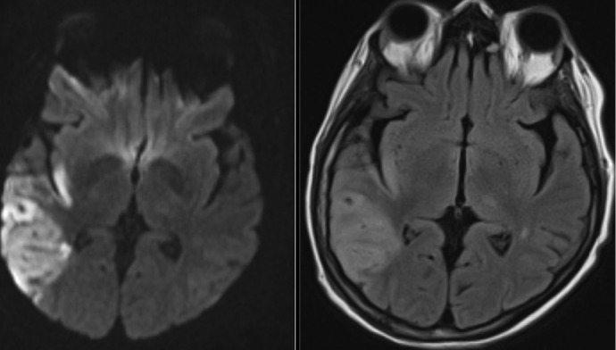
Figure 1.

Stroke complicating endocarditis. Axial diffusion-weighted imaging (left) and T2 fluid-attenuated inversion recovery (FLAIR) imaging (right) of a 64-year-old female with a history of severe mitral regurgitation who presented with confusion 2 weeks after a dental procedure. Imaging shows a large right middle cerebral artery territory embolic infarct. The patient was found to have Streptococcus mitis bacteremia and mitral valve endocarditis. Vessel imaging was patent, and she underwent successful valve repair 2 weeks after antibiotics were started.
