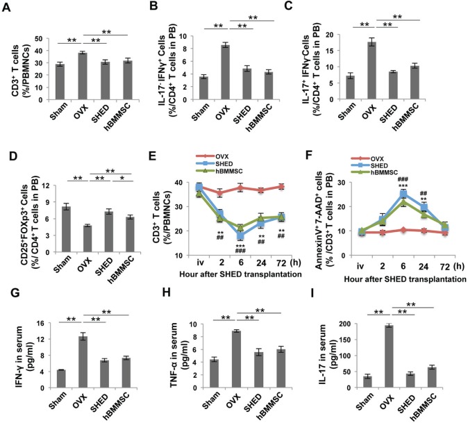
Figure 2.
SHED transplantation impeded changes in the T-cell subset in OVX mice. (A-D) Flow cytometric analysis showed that the percentage of CD3+ T-cells and the percentage of Th1 and Th17 cells in CD4+ T-cells were increased in peripheral blood of OVX mice. However, the percentage of Tregs in CD4+ T-cells was reduced. SHED and hBMMSC transplantation reduced the levels of CD3+ T-cells, Th1, and Th17 cells in OVX mice at 4 wk post-transplantation. SHED transplantation exhibited a stronger capacity to up-regulate the levels of Tregs in OVX mice compared with the hBMMSC transplantation group. (E, F) The percentage of CD3+ T-cells in peripheral blood was examined in a time-course by flow cytometric analysis. SHED or hBMMSC transplantation reduced the number of CD3+ T-cells and increased AnnexinV+7AAD+ apoptotic CD3+ T-cells in OVX mice, reaching a peak at 6 h. (G-I) ELISA analysis showed that OVX mice had elevated levels of IFN-γ, TNF-α, and IL-17 in peripheral blood when compared with the sham-operated control group. After SHED or hBMMSC transplantation, the levels of IFN-γ, TNF-α, and IL-17 were markedly reduced. N = 5 in each group; *p < .05; **p < .01; ***p < .005. Error bars: mean ± SD.

