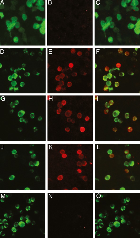
Figure 1.

Detection of LRP4 antibodies in sera of ALS patients using a cell-based assay (CBA). Immunofluorescence study of the binding of antibodies to HEK293 cells transfected with pCMV6-LRP4-tGFP or pEGFP. Left column: GFP fluorescence; middle column: bound antibody staining; right column: merged images Rows 1 and 2: Commercial rabbit LRP4 antibody (1:750 dilution) was incubated with pEGFP-transfected cells (row 1; A–C) or pCMV6-LRP4-tGFP-transfected cells (row 2; D–F) and bound antibodies visualized using Alexa 568-conjugated anti-rabbit IgG antibodies. Rows 3–5: LRP4-GFP-transfected cells were incubated with a 1:100 dilution of serum from two ALS patients (rows 3–4; G–L) or a healthy control (row 5, M–O) and bound antibodies visualized with Alexa 568-conjugated anti-human IgG antibodies. Only the commercial antibody and patients' sera bound on expressed LRP4 as visualized with red staining (E, H, K) and the merged images (F, I, L).
