
Figure 3.
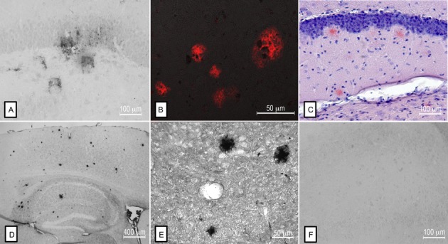
Comparison of Aβ deposition between SHRSP, Tg mouse models of AD, and human AD brains. Aβ-positive deposits in the hippocampus of a SHRSP (A) resemble those found in human AD brains (B). Similarly, plaques identified by Congo red staining (C) and by anti-Aβ immunohistochemistry (D, E) in mice overexpressing the Swedish human APP mutation, have a similar morphology to those identified in the SHRSP. Preabsorption of anti-Aβ (clone 4G8) with human Aβ40 attenuated the detection of Aβ staining in SHRSP (F).