
Fig. 2. Paddlefish Pax3 is expressed in the profundal placode and ganglion.
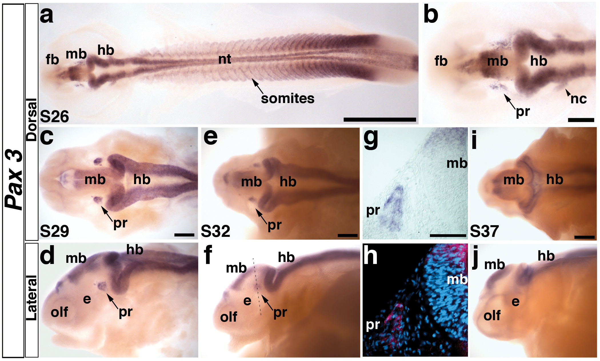
(a) Embryo at stage 26 showing the distribution of Pax3 transcripts in the neural tube, somites, and head. At this stage, the primary divisions of the head into forebrain (fb), midbrain (mb) and hindbrain (hb) are evident. (b) Higher power view of head region in panel a. Pax3 is expressed in bilateral patches of cells lateral to the midbrain (mb) in the profundal placode (pr) and in neural crest cells (nc) migrating from the hindbrain (hb) towards the pharyngeal arches. (c,d) Stage 29 embryo in dorsal and lateral views, respectively. The Pax3-positive cells in the profundal placode (pr) have condensed, yet remain near the epithelium. Pax3 is also weakly expressed in the olfactory pits (olf). (e,f) Stage 32 embryo in dorsal and lateral views. The Pax3-positive cells have delaminated from the epithelium into the mesenchyme in the developing profundal ganglion (pr). Dotted line shows plane of section shown in panels g and h. (g) Brightfield image of a transverse section through the stage 32 embryo shown in panel f. Pax3-positive cells are clearly expressed in the developing profundal ganglion (pr). (h) Corresponding false color overlay image of brightfield image in panel g (red, Pax3) with the nuclear marker DAPI (blue). (i,j) Stage 37 embryo in dorsal and lateral views. Pax3 expression persists in the central nervous system, yet is completely absent in the profundal ganglion by this stage. Abbreviations: e, eye; fb, forebrain hb, hindbrain; mb, midbrain; nt, neural tube; olf, olfactory pits; pr, profundal placode or ganglion; s, stage. Scale bars: (a) 1 mm; (b-f, i, j) 200 μm; (g,h) 50 μm.