
Fig. 5. Paddlefish Phox2b is expressed in epibranchial placode-derived neurons.
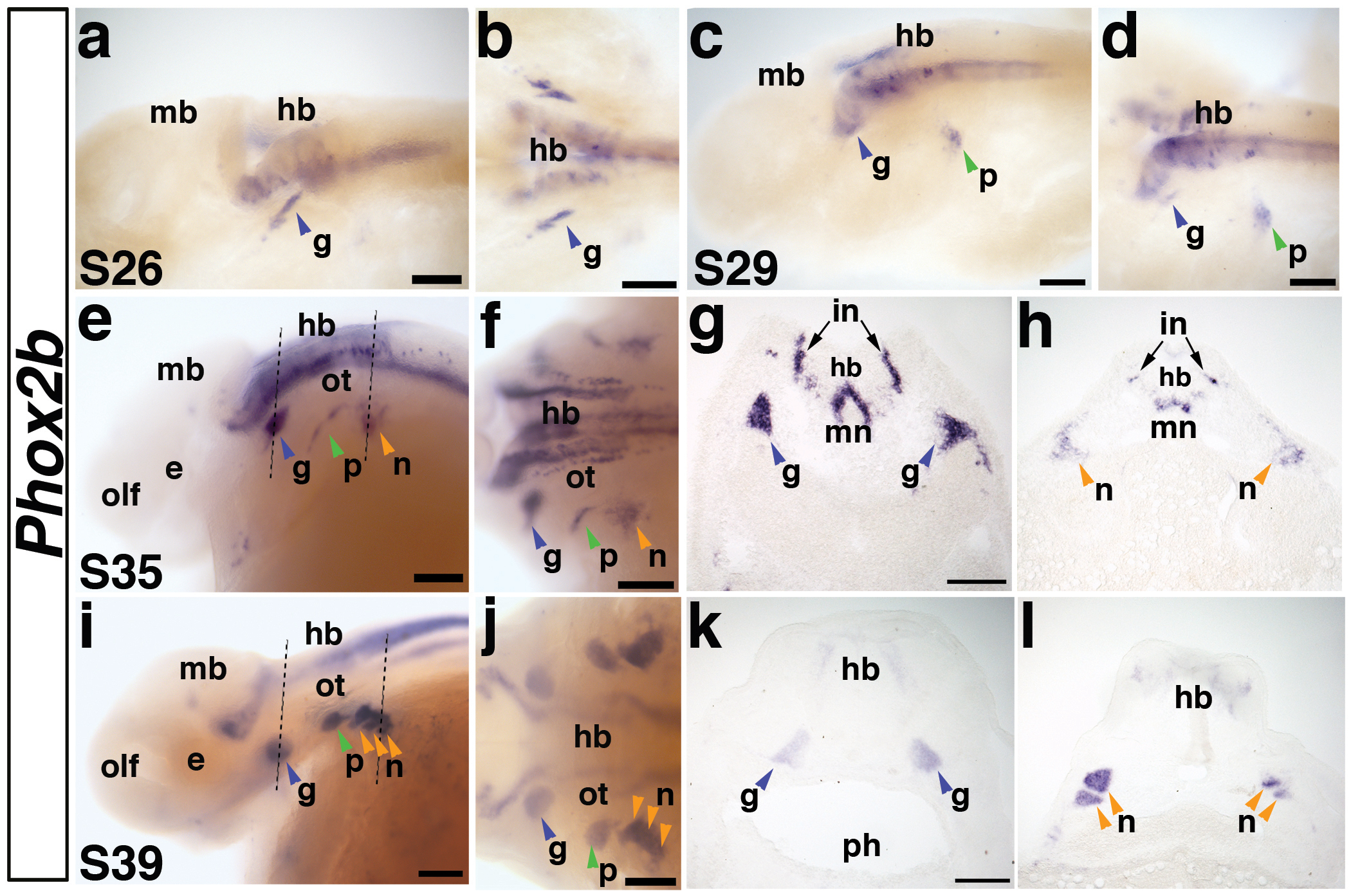
(a,b) Lateral and dorsal views, respectively, of a stage 26 embryo. Phox2b is expressed in a stripe of cells representing the first neurons of the developing geniculate ganglion (g) (blue arrowhead) and in neurons in the developing hindbrain (hb). Expression of Phox2b in the hindbrain (hb) persists throughout embryogenesis. (c,d) Lateral and dorsal views of a stage 29 embryo. Phox2b is expressed in the condensing geniculate ganglion (blue arrowhead) and now also in the developing petrosal ganglion (p) (green arrowhead). (e,f) Lateral and dorsal views of a stage 35 embryo. Phox2b expression continues in the geniculate ganglion (blue arrowhead), petrosal ganglion (green arrowhead), and has begun in a patch of cells representing the developing nodose ganglia (n) (orange arrowhead). Dotted lines in panel e indicate planes of sections shown in panels g and h. (g) Section through the geniculate ganglia (blue arrowheads) of the stage 35 embryo shown in panels e,f. Phox2b transcripts are strongly expressed in the geniculate ganglia, as well as in a subset of hindbrain neurons, presumably including interneurons (in) and motor neurons (mn). (h) Section through the developing nodose ganglia (orange arrow) in the stage 35 embryo shown in panel e. Compared to the condensed geniculate ganglion, Phox2b-positive cells of the developing nodose ganglia are still scattered, such that individual ganglia cannot be distinguished. (i,j) Lateral and dorsal views of a stage 39 embryo. Phox2b expression clearly marks all the condensed epibranchial placode-derived ganglia (arrowheads). Dotted lines in panel i indicate planes of sections in panels k and l. (k) Section through the geniculate ganglia (blue arrowheads) of the stage 39 embryo shown in panels i,j, showing Phox2b expression in the medially located ganglia. (l) Section through the nodose ganglia (orange arrowheads) of the stage 39 embryo shown in panels i,j. At this stage, individual Phox2b-positive nodose ganglia can be distinguished. Abbreviations: e, eye; g, geniculate ganglion; hb, hindbrain; in, interneurons; mb, midbrain; mn, motor neurons; n, nodose ganglia, olf, olfactory pits; ot, otic; p, petrosal ganglion; ph, pharynx; s, stage. Scale bars: (a-f; i-j) 200 μm; (g,h) and (k,l) 100 μm.