
Fig. 6. Paddlefish Sox3 is expressed in epibranchial and lateral line placodes and sensory organs.
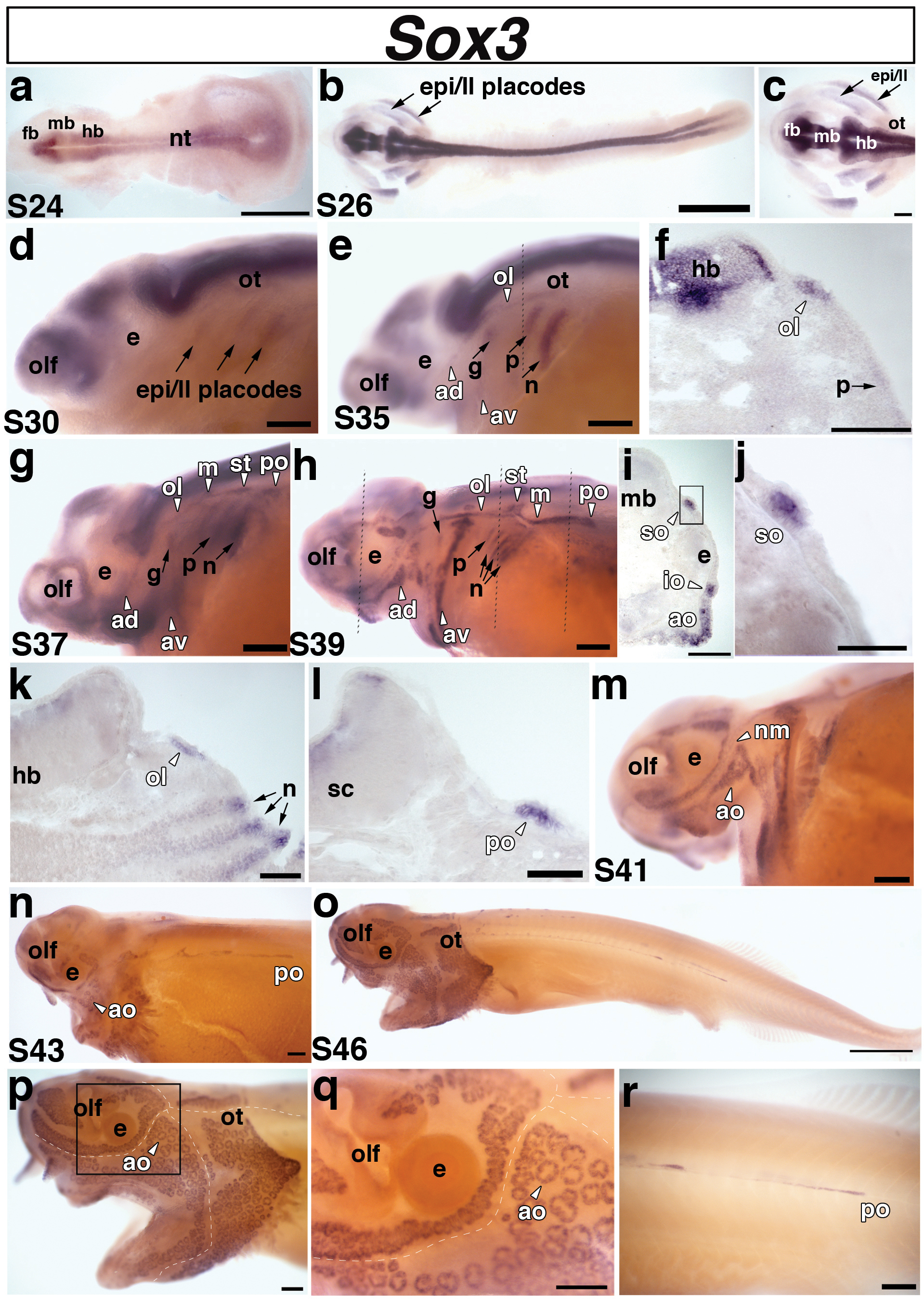
Lateral views unless stated otherwise. (a) Dorsal view of a stage 24 embryo. Sox3 is expressed in the neural tube. (b) Dorsal view of a stage 26 embryo and (c) higher power view of the head region of the stage 26 embryo shown in panel b. Sox3 is expressed in two broad domains representing the presumptive epibranchial and lateral line placodes (arrows) in the pharyngeal arch region. (d) Stage 30 embryo. Sox3 is still present, although faintly, in the lateral regions that comprise the epibranchial and lateral line placodes (epi/ll, arrows). (e) Stage 35 embryo. At this stage, Sox3 can be distinguished in the different epibranchial placodes: the geniculate (g), petrosal (p), and nodose (n) (arrows), and in the pre-otic lateral line placodes: anterodorsal (av), anteroventral (av), otic lateral line (ol) (arrowheads). The lateral line placodes have begun to elongate and form sensory ridges in the head. Sox3 is also expressed in the olfactory pits (olf). Dotted line shows plane of section shown in panel f. (f) A section through the hindbrain (hb) of the embryo shown in panel e reveals separate domains of Sox3 expression in the otic lateral line placode (ol) and faintly in the petrosal placode (p). (g) Stage 37 embryo. Sox3 persists in the epibranchial (arrows) and elongating lateral line (arrowheads) placodes. At this stage, Sox3 appears to be expressed in all six lateral line placodes: anterodorsal (av), anteroventral (av), otic lateral line (ol), middle (m), supratemporal (st), and posterior (po). (h) Stage 39 embryo. Sox3 expression persists in the elongating lateral line primordia, but the boundaries of individual primordia become difficult to determine owing to the large size of some of the ridges. At this stage, the posterior lateral line placode (po) has begun its extensive migration posteriorly. Sox3 also continues to be expressed in the epibranchial placodes, although in a weaker, more restricted dorsal region. Dotted lines show planes of sections shown in panels i-l. (i-l) Sections from different rostrocaudal positions of the stage 39 embryo shown in panel h. (i) Section through the eye. Sox3 transcripts are clearly expressed in the developing supraorbital (so) and infraorbital (io) neuromast (mechanosensory) lines. A broad patch of Sox3 expression can also been seen in a developing field of electrosensory ampullary organs (ao) ventral to the infraorbital (io) canal. (j) Higher power view of boxed area in panel i, showing Sox3 transcripts in the supraorbital (so) neuromast canal line. (k) Section through the hindbrain. Sox3 is expressed in the otic lateral line placode (ol) (arrowhead) and in the nodose (n) placodes (arrows). It is also expressed more weakly in “stripes” of cells subjacent to the nodose placodes that may represent pharyngeal pouch endoderm. (l) Section through the spinal cord. Sox3 is seen in the posterior lateral line primordium (po) of the main trunk line. (m) Stage 41 embryo. Sox3 expression is maintained in both mechanosensory neuromasts (nm) and electrosensory ampullary organs (ao), but is no longer seen in the epibranchial placodes. (n) Stage 43 embryo. Sox3 expression is maintained in the developing ampullary organ (ao) fields. The lines of neuromasts in the head are already recessed within lateral line canals and Sox3 expression is no longer visible in these lines, but can still be seen in the migrating posterior lateral line primordium (po). (o-r) Stage 46 embryo. (o,p) Sox3 is maintained in distinct fields of ampullary organs on the head. (q) Higher power view of the boxed area in panel p. Sox3 can be seen in individual clusters of ampullary organs. In panels p and q, the positions of the head neuromast canal lines are indicated by the dotted white lines. (r) Higher power view of the migrating Sox3-positive posterior lateral line placode (po). Abbreviations: ad, anterodorsal lateral line placode; ao, ampullary organs (electroreceptors); av, anteroventral lateral line placode; e, eye; epi, epibranchial; fb, forebrain; hb, hindbrain; in, interneurons; io, infraorbital neuromast canal; ll, lateral line; m, middle lateral line placode; mb, midbrain; nm, neuromasts; nt, neural tube; ol, otic lateral line placode; olf, olfactory pits; ot, otic; po, posterior lateral line placode of the main trunk; s, stage; so, supraorbital neuromast canal; st, supratemporal lateral line placode. Scale bars: (a,b;o) 1 mm; (c-e; g,h; m,n; p-r); 200 μm (f,i,k) 100 μm; (j,l) 50 μm.