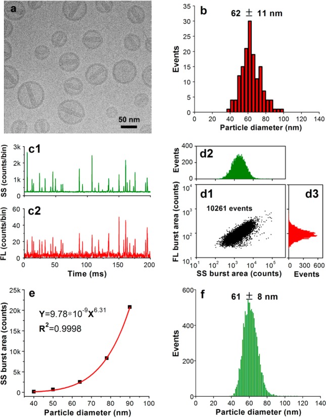
Figure 4.
Characterization of doxorubicin-encapsulating liposomes. (a) Representative cryo-TEM image of Doxoves. (b) Particle size distribution histogram of Doxoves liposomes obtained from cryo-TEM images. (c) Representative SS and FL burst traces of Doxoves (diluted 4000-fold in 5% glucose) measured using HSFCM. (d) Bivariate dot-plot of the FL versus the SS burst area for Doxoves preparation. (e) Plot of nanoparticle SS burst area as a function of particle size for monodisperse silica nanoparticle standards. (f) Particle size distribution profile of Doxoves liposomes measured using HSFCM and the calibration curve presented in panel e.

