Figure 4.
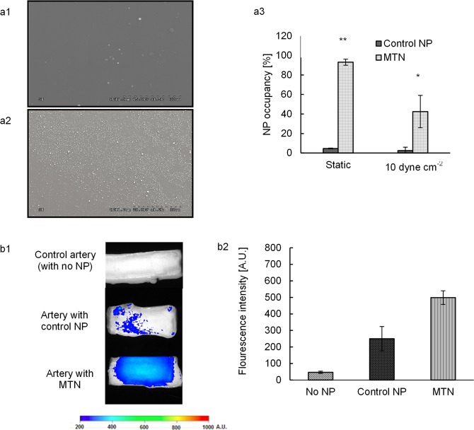
In vitro and ex vivo targeting efficiency of MTNs. (a1, a2) Scanning electron microscopy images of control NP and MTN adhering on vWF-coated surfaces, respectively, under 10 dyn/cm2. (a3) Quantification of surface coverage by particle adhesion via ImageJ. (b1) Fluorescent images of arteries with lumen injury after incubating with control particles and MTNs. (b2) The fluorescent intensity was determined and compared for three groups: control (no NP), control NP (unconjugated NPs), and MTNs. * represents p < 0.05, whereas ** represents p < 0.01. N = 5.

