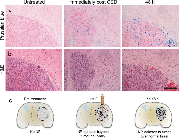
Figure 6.
NPCP-BG-CTX is preferentially retained in the tumor region. (a) Representative Prussian blue and (b) H&E stained subsequent tissue sections of brain at the tumor margin obtained from untreated or NPCP-BG-CTX treated animals immediately post CED or 48 h post CED. The scale bar corresponds to 100 μm. (c) Schematic illustration of time dependent changes of NP localization before and after CED.

