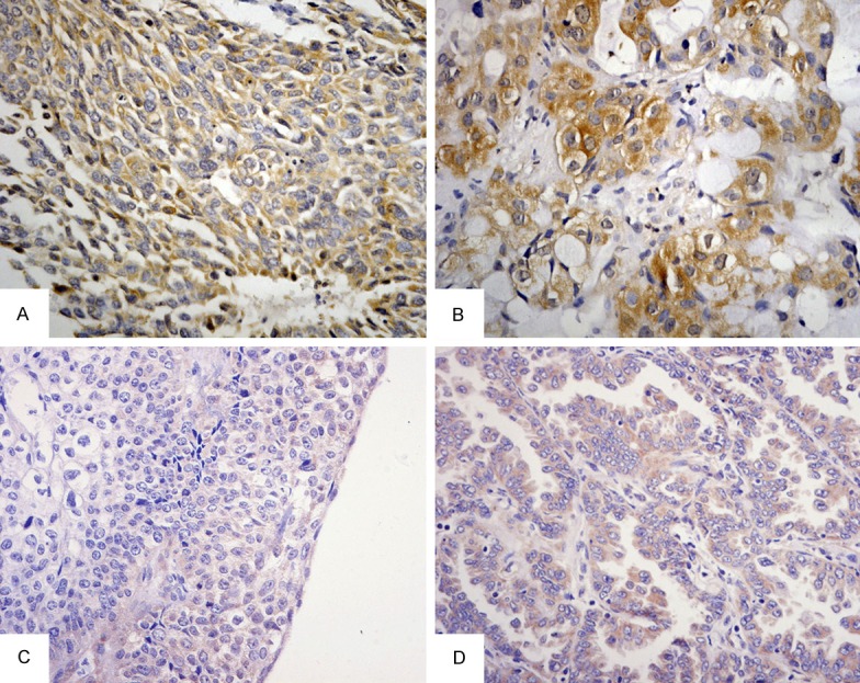
Figure 1.

IHC and ISH of annexin A1 in lung cancer and adjacent-cancer normal tissues (×400). A. high staining of annexin A1 in poorly differential LSCC; B. high staining of annexin A1 in poorly differential LAC; C. Low staining of annexin A1 mRNA in poorly differential LSCC; D. Low staining of annexin A1 mRNA in well differentiated LAC. LSCC, squamous cell carcinoma of the lung; LAC, adenocarcinoma of the lung.
