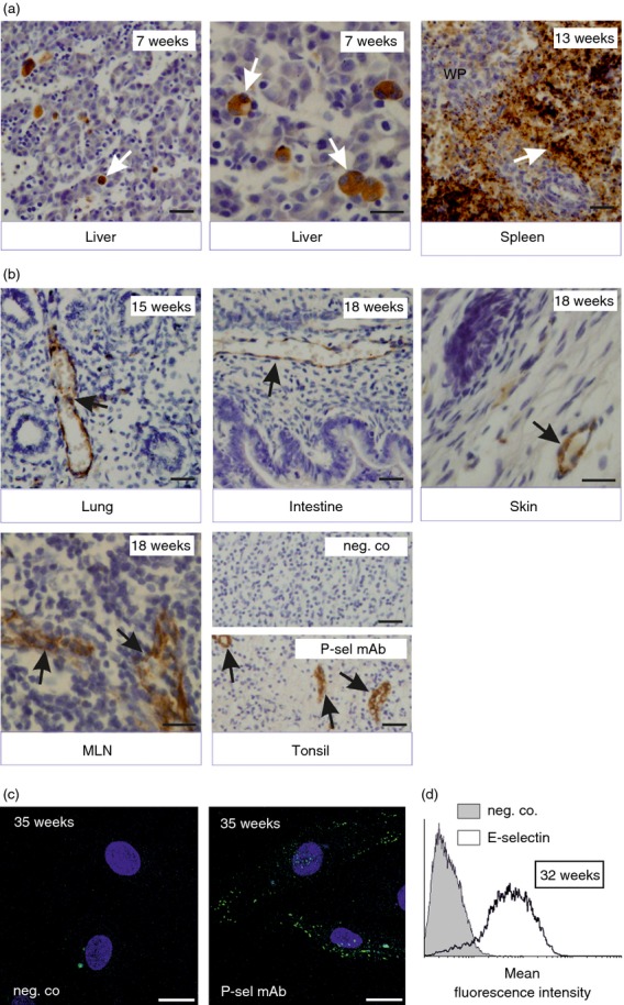
Figure 1.

Endothelial selectins are expressed during fetal development in humans. (a–b) Expression of P-selectin in (a) megakaryocytes and platelets and (b) vascular endothelium in fetal organs. Gestational age is indicated, and white arrows point to megakaryocytes and platelets and black arrows point to vessels; WP, white pulp. Scale bar, 50 μm. (c, d) Human umbilical vein endothelial cells (HUVEC) isolated from preterm deliveries were (c) left untreated and stained for negative control and P-selectin and analysed by microscopy (blue is DAPI, scale bar, 20 μm) or (d) stimulated with tumour necrosis factor-α for 4 hr and stained for E-selectin using FACS. Representative data from at least three independent experiments with different donors are shown.
