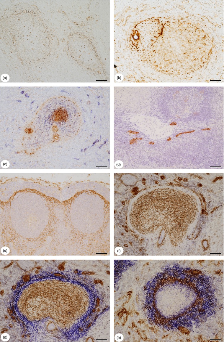
Figure 2.

(a) Distribution of CXCL12 in the human splenic white pulp. Cells and/or fibres surrounding periarterial lymphatic sheath (PALS) and follicles are positive. (b) Distribution of CXCL13 in the human splenic white pulp. A small mixed periarterial region or a PALS (left) with a follicle (right) are surrounded by CXCL13+ stromal cells and/or fibres. Follicular dendritic cells (FDCs) in the mantle zone of the follicle are weakly positive. Smooth muscle cells and adventitial stromal cells or fibres surrounding an arteriole (left) are also stained. (c) Double-staining for CXCL13 (brown) and CD271 (blue) in a small periarterial region or PALS (left) and a secondary follicle (right). The FDCs in the germinal centre are strongly CXCL13+, whereas FDCs in the mantle zone and cells and/or fibres at the surface of the periarterial region and the follicle stain more weakly. Adventitial stromal cells or fibres and smooth muscle cells of arterial walls are also CXCL13+. (d) Distribution of podoplanin in the adult human splenic white pulp. Podoplanin is only visualized in endothelia of a lymphatic vessel network accompanying a large artery. Fibroblastic reticulum cells (FRCs) are not stained. Single ABC-DAB staining with monoclonal antibody 4D5aE5E6 and haemalum nuclear counterstain. (e) Expression of receptor activator of nuclear factor-κB ligand (RANKL) (CD254) in a lymph node situated at the splenic hilum (most probably a gastric lymph node). RANKL is most strongly expressed in branched stromal cells of the subcapsular sinus floor and more weakly in stromal cells surrounding two secondary follicles. Single staining for RANKL with polyclonal antibody and haemalum nuclear counterstain. (f) Control section of a follicle stained for CD271 (brown) with omission of the second primary antibody (mucosal addressin cell adhesion molecule-1; MAdCAM-1), but with Ultravision and AP-reaction. The CD271− region is especially well visible, because of weak background staining for endogenous AP in perifollicular cells. (g) Double-staining for CD271 (brown) and MAdCAM-1 (blue) in a follicle. The section shows the same follicle as (f). The CD271+ FDCs (brown) and the MAdCAM-1+ CD271− superficial stromal cells join one another without any unstained area in between. As far as recognizable after subtractive staining, the two stromal cell types do not overlap at the border of the mantle zone. The round or elongated dark brown structures are capillary sheaths. (h) Double-staining for SMA (brown) and MAdCAM-1 (blue) at the follicular surface. In this patient the staining for MAdCAM-1 is much more widespread than that for SMA. MAdCAM-1 stains many stromal cells located outside the SMA+ shell, but also some stromal cells located inside. (a, d, f–h) 17-year-old male patient, (b, c) 22-year-old male patient, (e) 15-year-old male patient. All patients had suffered an extraabdominal trauma. (b, c) Paraffin sections, (a, d–h) cryosections. (a, b, d, e) Single ABC-DAB staining. (c, g, h) ABC-DAB staining for first primary antibody followed by Ultravision-AP with Fast Blue chromogen for second primary antibody. (f) Omission of second primary antibody. Scale bars: a, c–h = 100 μm, b = 50 μm.
