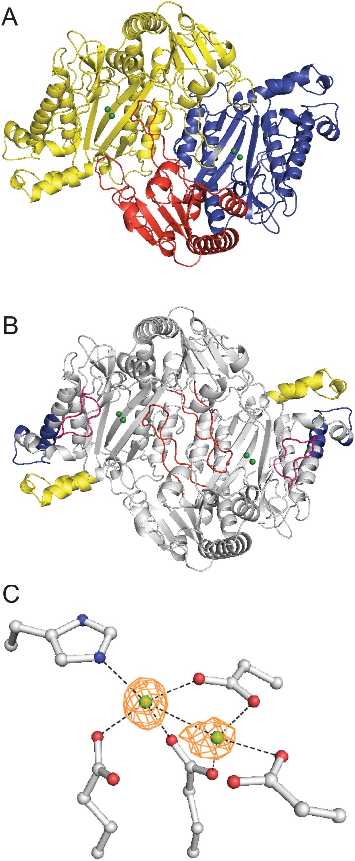
Figure 2. PepQ forms a canonical pita-bread fold with binuclear active site.
(A) The PepQ dimer (PDB entry 4QR8) is shown with one monomer shown in yellow and one monomer colored by domain: N-terminal (residues 1–159, red) and catalytic (160–443, blue). The magnesium ions are colored green. The image was rendered in PyMOL [63]. (B) The PepQ dimer with new regions of sequence (those not in P. furiosus) highlighted (residues 35–53, red; 303–321, blue; 360–372, pink; 391–415, yellow). (C) Electron density shows conserved active site residues coordinating two magnesium ions.

