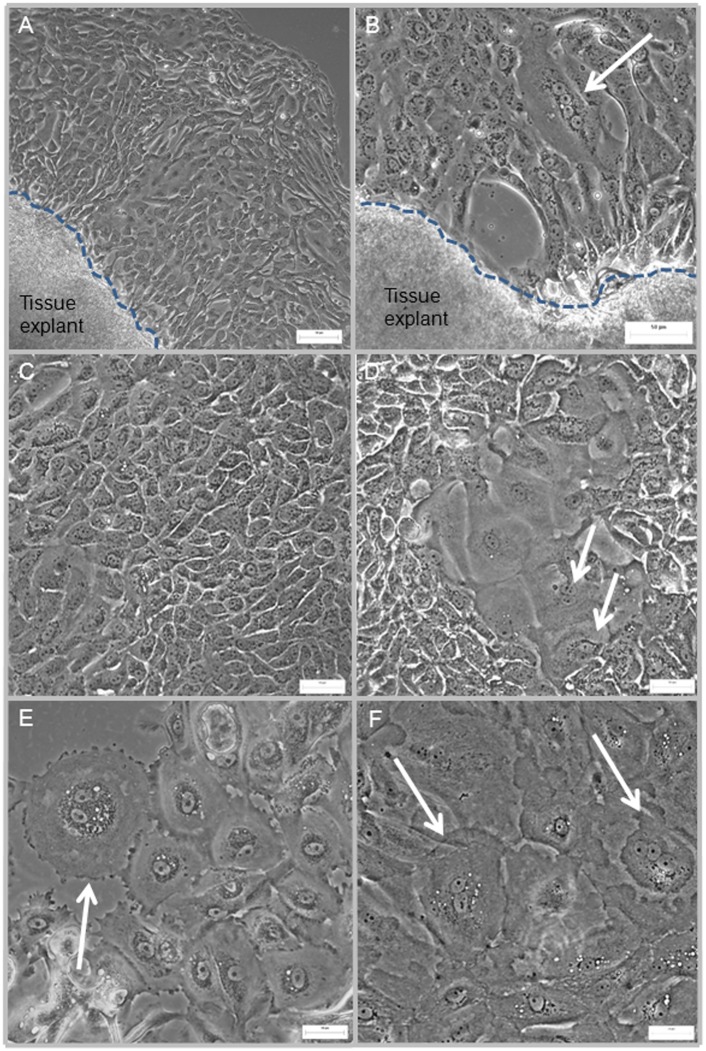
Figure 1. Cellular morphology of human urothelial cells (HUCs) generated from bladder tissue explant.
(A, B) Cells growing out from the edge of two tissue explants obtained from two different biopsies; cells with typical coble-stone morphology were clearly identifiable. Arrow points to a larger cell (60–150 µm) with four nuclei and prominent nucleolus. (C, D) cells with uniform size and morphology (25–40 µm) surround a cluster of larger cells (70–100 µm); several are bi-nucleated (arrows). (E, F) clusters of larger cells (80–150 µm), many of these cells are bi-nucleated (arrows). The dotted lines demarcate the edge of the tissue explants. Scale bar = 50 µm.

