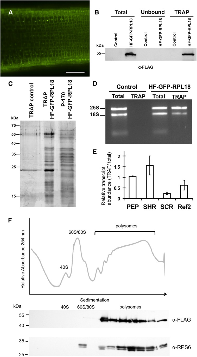
Figure 5.
Application of TRAP methodology to tomato with the hairy root transformation system. A, 35Spro-driven His6-FLAG epitope-tagged GFP-RPL18 (HF-GFP-RPL18) fusion protein in the tomato hairy root system shows GFP localization (green) in the nucleolus and cytosol. Bar = 100 μm. B, HF-GFP-RPL18 is efficiently immunopurified by TRAP. Equal fresh weight of tissue from wild-type roots (control) and HF-GFP-RPL18 hairy roots was extracted in a polysome-stabilizing buffer and processed to obtain a clarified cell supernatant (total), which was incubated with anti-FLAG agarose beads to obtain the TRAP fraction containing ribosomes and associated mRNA. Cell components not bound to the matrix remained in the unbound fraction. Molecular mass markers are shown on the left. The expected size of HF-GFP-RPL18 is 51 kD. C, Comparison of immunopurified proteins from untransformed roots or HF-GFP-RPL18-transformed hairy roots by TRAP and ribosomal proteins isolated by conventional ultracentrifugation (P-170). Proteins were visualized by silver staining. Equal proportions of TRAP samples were loaded in lanes 1 and 2. Molecular mass markers are shown on the left. Asterisks indicate bands corresponding to IgG from the affinity matrix. D, Ethidium bromide staining of RNA isolated from the total and TRAP fractions from wild-type (control) and HF-GFP-RPL18 roots. The 25S and 18S rRNA sizes are indicated on the left. E, Quantitative reverse transcription-PCR analysis of selected transcripts in the TRAP fractions. Relative transcript level in the TRAP versus total fraction was determined. Data represent average ± sd level of each transcript in two biological replicates. ACT2 was used as a reference for normalization. F, HF-GFP-RPL18 is incorporated into small to large polysomal complexes. HF-GFP-RPL18 hairy root ribosomal complexes were fractionated by ultracentrifugation through a 15% to 60% Suc density gradient, and the A254 was recorded. The positions of ribosomal subunits 40S and 60S, monosomes (80S), and polysomes are indicated. Fourteen fractions were analyzed by immunoblot with anti-FLAG and anti-RPS6 antisera. Two electrophoretic variants of RPS6 were detected by the antisera. Molecular mass markers are indicated on the left.

