Figure 5.
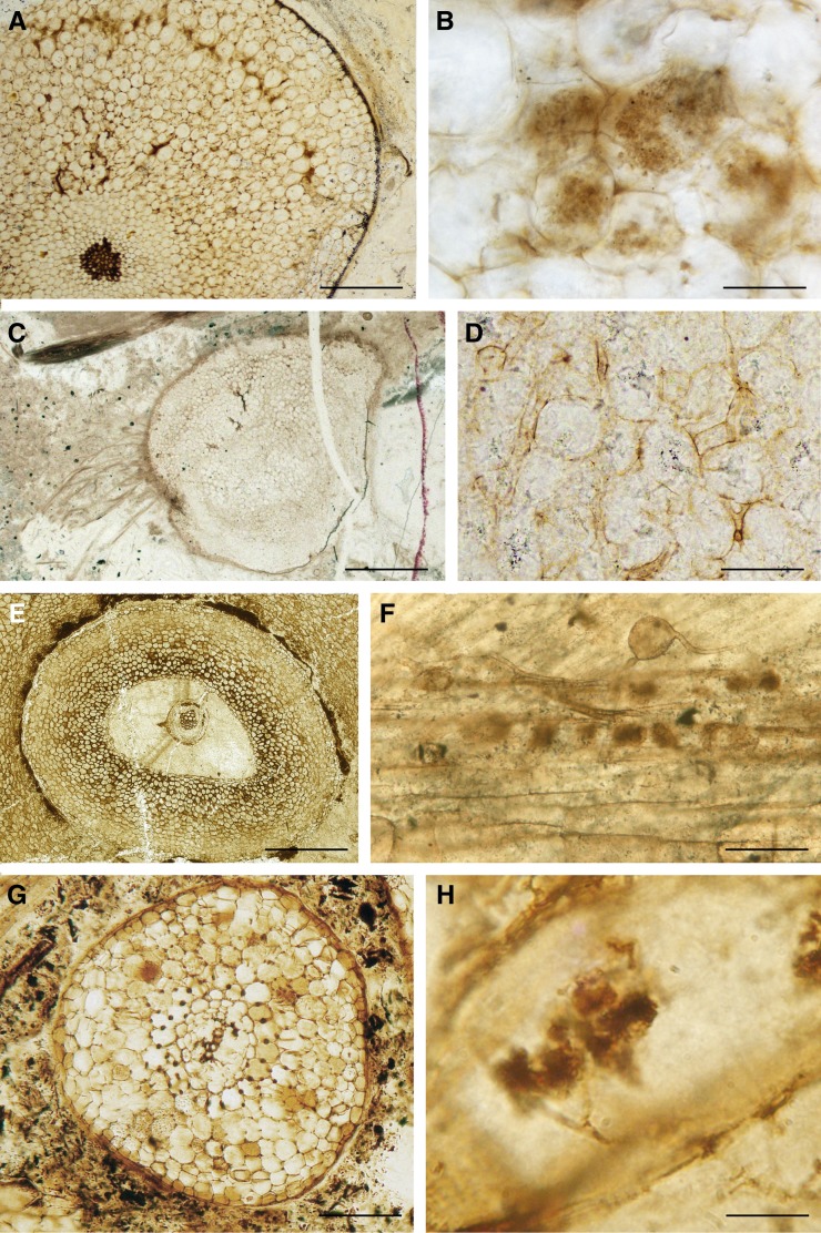
Fungal partnerships in Devonian and Carboniferous plants. A and B, Fungal endophyte of the glomeromycotan type in A. major from the Devonian Rhynie Chert. A, Transverse section of an aerial axis showing the well-defined colonized zone in the outer cortex (slide PB V15637 from the Natural History Museum, London). B, Arbuscule-like structures in an aerial axis (slide from the University of Munster; photograph courtesy of H. Kerp). C and D, Colonization of the mucoromycotean type in H. lignieri from the Devonian Rhynie Chert. C, Transverse section of a corm; a zonation of fungal colonization is visible within the corm. D, Intercellular branched thin-walled and intercellular thick-walled hyphae are present. E, Arborescent clubmoss rootlet from the Upper Carboniferous of Great Britain (slide PB V11472 from the Natural History Museum, London). F, AM-like fungi in stigmarian appendage. Trunk hyphae, intercalary vesicle (left), and putative arbuscule-like structures (right) are visible (slide BSPG 1964X from the Bavarian State Collection for Paleontology and Geology; photograph courtesy of M. Krings). G, Cordaites rootlet from the Upper Carboniferous of Grand’Croix, France, colonized by AM fungus. The cortex comprises a reticulum of phi thickenings that are prominent in cells located close to the vascular cylinder (slide Lignier Collection no. 194 from the University of Caen). H, Detail of an arbuscule-like structure. The hyphal trunk of the arbuscule-like structure branches repeatedly forming a bush-like tuft within the cell (slide Lignier Collection no. 194 from the University of Caen). Bars = 0.55 mm in A, 30 µm in B, 1.1 mm in C, 120 µm in D, 1.5 mm in E, 70 µm in F, 1.25 mm in G, and 18 µm in H.

