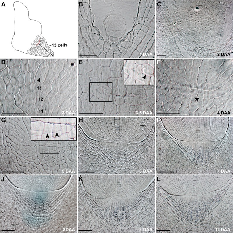
Figure 1.
Origin and development of the root cap during rice embryogenesis. A, Schematic view of a medial longitudinal section through a rice embryo at around 4 DAA. The red line denotes the developing cap junction. The black line shows an average 13-cell distance between the cell at the basal end of the suspensor and the developing cap junction. B, Embryo at 1 DAA. C, Embryo at 2 DAA. D to F, Cells at the region where the radicle and cap junction originate. The 11, 12, and 13 in D denote the cell counts at the distance from the cell at the basal end of the suspensor of an embryo at 3.5 DAA. The inset in E is an enlarged view of the boxed region. Arrowheads in D to F point to the putative initiation position of the cap junction (D), an extending part of the cap junction formed by an anticlinal cell division (E), and a daughter cell produced by an anticlinal division of the root cap stem cell (F), respectively. G to L, Development of the root cap in the embryo at 5 DAA (G), 6 DAA (H), 7 DAA (I), 8 DAA (J), 9 DAA (K), or 12 DAA (L). The inset in G is an enlarged view of the boxed region. Arrowheads in G indicate that more root cap stem cells divided at 5 DAA. Lugol staining showed that starch granules appeared from 7 DAA in the lower 10 layers of columella root cap cells (I) and starch granules in lateral root cap cells could only be readily seen from 8 DAA (J–L). Scale bar = 25 μm for B to F and 50 μm for G to L.

