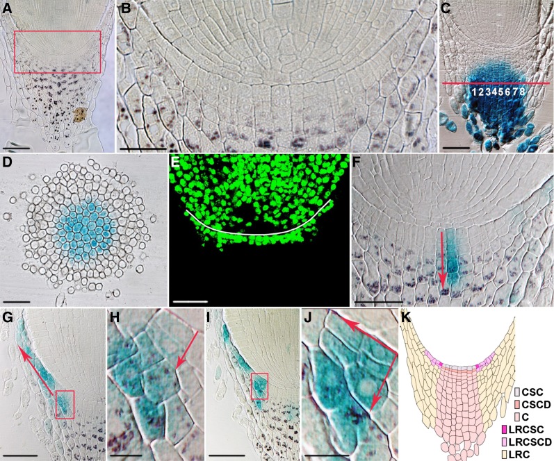
Figure 2.
Cell fate and cell lineage in the radicle root cap. A, Medial longitudinal section of the radicle root tip. Starch granules in the columella root cap were revealed by Lugol staining. B, Enlarged view of the boxed region in A. C, Medial longitudinal section of the radicle root tip of the columella root cap-specific GAL4/UAS::GUS enhancer trap line A788. Note that GUS staining could be observed in eight columns of columella root cap cells. D, Cross section at the position indicated by the red line in C. Cells with GUS staining are columella root cap cells, surrounded by three to five layers of GUS-negative lateral root cap cells. E, EdU cell proliferation assay revealing stem cells and their daughters in the root cap. The white line denotes the position of the cap junction. F to J, Medial longitudinal sections of the radicle root tips of the 35S::Spm-GUS lines, stained with both GUS and Lugol solution. GUS-marked clonal sectors suggest the presence of both columella (F) and lateral root cap lineages (G–J). Arrows in F to H and J show the direction of cell division. H and J are enlarged views of the boxed regions in G and I, respectively. K, Cartoon showing the medial longitudinal view of the root cap. Cell types are color coded. C, Columella; CSC, columella stem cell; CSCD, columella stem cell daughter; LRC, lateral root cap; LRCSC, lateral root cap stem cell; LRCSCD, lateral root cap stem cell daughter. All images were taken at the same magnification except B, H, and J. Bar = 50 μm for A, C to G, and I; 25 μm for B; and 10 μm for H and J.

