Figure 3.
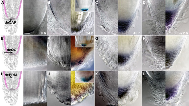
Root cap regeneration after deCAP, deQC, or dePRM. A to D, Morphology of the radicle root tips at 0 h (A, right), 24 h (B), 48 h (C), or 72 h (D) after deCAP, without (A, right; and B–D, left) or with (B–D, right) Lugol staining. The left image in A is a cartoon showing the medial longitudinal view of the radicle root tip. The arrow points to the position of deCAP. The inset in B shows that GUS expression was detected in A788 24 h after deCAP. E to H, Morphology of the radicle root tips at 0 h (E, right), 24 h (F), 48 h (G), or 72 h (H) after deQC, without (E, right; and F–H, left) or with (F–H, right) Lugol staining. The arrow in E (left) points to the position of deQC. The inset in F shows that GUS expression was detected in A788 24 h after deQC. I to L, Morphology of the radicle root tips at 0 h (I, right), 24 h (J), 48 h (K), or 72 h (L) after dePRM, without (I, right; and J–L, left) or with (J–L, right) Lugol staining. The arrow in I (left) points to the position of dePRM. The inset in J shows that GUS expression was detected in A788 24 h after dePRM. All images were taken at the same magnification. Bar = 50 μm.

