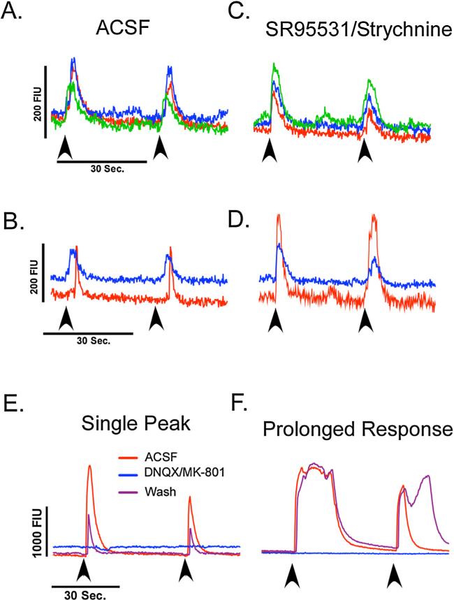
Fig. 5.
(A) Representative traces from three cells recorded simultaneously demonstrating a time-locked increase in fluorescent activity following electrical stimulation of the NST in normal ACSF. (B) Representative trace of cells in the same field that demonstrated a delayed response to electrical stimulation (red trace) and time-locked response (blue trace). Arrows indicate the stimulation onset. (C) When GABAA and glycine receptors were blocked with SR95531 and strychnine (5 μM), no change was seen in cells that had time-locked responses. (D) The cell that had a delayed response however, became time-locked to the stimulus and had an increased amplitude in fluorescent intensity. (E and F) Representative traces of cells in the cNST that were activated by rNST stimulation (red trace). Ionotropic glutamate receptor antagonists (DNQX and MK-801, 10 μM) abolished the increases in fluorescent intensity (blue trace) that recovered following washout (purple trace). (For interpretation of the references to color in this figure legend, the reader is referred to the web version of the article.)

