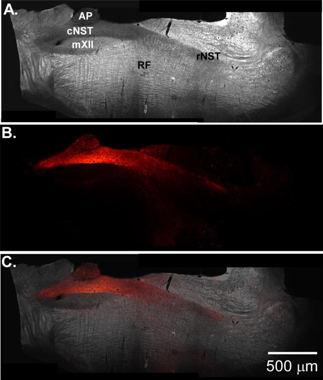
Fig. 7.
(A) Dark-field photomicrograph of oblique section from rat pup injected with an adeno-associated viral vector that expresses a channelrhodopsin-2(h134r)-mCherry fusion protein under the control of the synthetic promoter PRSx8 (Nasse and Travers 2013). (B) Fluorescent photomicrograph of the same slice showing transfected cells in the cNST and fibers projecting to the rNST and reticular formation. (C) Overlay of (A) and (B).

