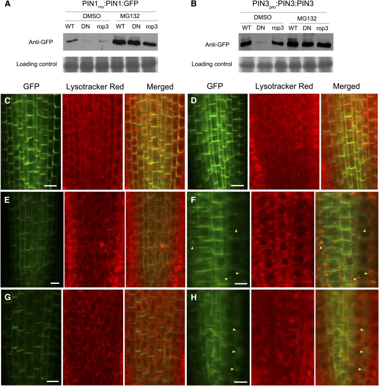
Figure 11.
PIN1:GFP Partially Accumulates in Vacuoles in MG132-Treated Roots of 35Spro:DN-rop3 and rop3.
(A) and (B) PIN1: GFP (A) and PIN3:GFP (B) protein levels in MG132-treated and untreated wild-type, DN-rop3, and rop3 seedlings. DN represents 35Spro:DN-rop3. Immunoblot was performed using anti-GFP antibody. Lower panels indicate Coomassie blue staining of the protein samples as loading controls
(C) and (D) PIN1:GFP localization in roots of 5-d-old seedlings not treated (C) or treated with MG132 (D).
(E) and (F) PIN1:GFP localization in roots of 35Spro:DN-rop3 seedlings not treated (C) or treated with MG132 (D).
(G) and (H) PIN1:GFP localization in roots of rop3 seedlings untreated (E) or treated with MG132 (F).
Lysotracker red was used for labeling of vacuolar compartments. Arrowheads mark the vacuoles containing PIN1:GFP. Bars = 10 μm.

